Deposition Date
1997-07-30
Release Date
1998-08-05
Last Version Date
2024-10-30
Entry Detail
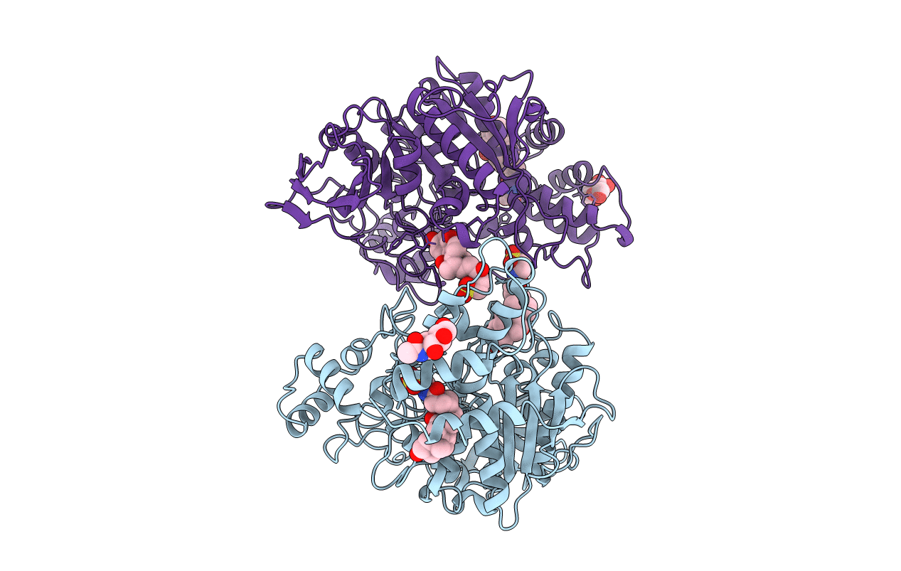
PDB ID:
1AQL
Keywords:
Title:
CRYSTAL STRUCTURE OF BOVINE BILE-SALT ACTIVATED LIPASE COMPLEXED WITH TAUROCHOLATE
Biological Source:
Source Organism(s):
Bos taurus (Taxon ID: 9913)
Method Details:
Experimental Method:
Resolution:
2.80 Å
R-Value Free:
0.27
R-Value Work:
0.21
R-Value Observed:
0.21
Space Group:
P 21 21 2