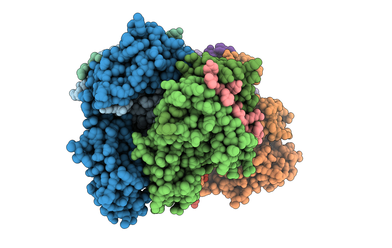
1AQD image
Entry Detail
PDB ID:
1AQD
Title:
HLA-DR1 (DRA, DRB1 0101) HUMAN CLASS II HISTOCOMPATIBILITY PROTEIN (EXTRACELLULAR DOMAIN) COMPLEXED WITH ENDOGENOUS PEPTIDE
Biological Source:
Source Organism:
Host Organism:
PDB Version:
Deposition Date:
1997-07-28
Release Date:
1998-01-28
Method Details:
Experimental Method:
Resolution:
2.45 Å
R-Value Free:
0.27
R-Value Work:
0.21
R-Value Observed:
0.21
Space Group:
C 1 2 1
Macromolecular Entities
Polymer Type:polypeptide(L)
Description:HLA-DR1 CLASS II HISTOCOMPATIBILITY PROTEIN
Chain IDs:A, D, G, J
Chain Length:192
Number of Molecules:4
Biological Source:Homo sapiens
Polymer Type:polypeptide(L)
Description:HLA-DR1 CLASS II HISTOCOMPATIBILITY PROTEIN
Chain IDs:B, E, H, K
Chain Length:198
Number of Molecules:4
Biological Source:Homo sapiens
Polymer Type:polypeptide(L)
Description:HLA-A2
Chain IDs:C, F, I, L
Chain Length:15
Number of Molecules:4
Biological Source:Homo sapiens
Primary Citation
The class II MHC protein HLA-DR1 in complex with an endogenous peptide: implications for the structural basis of the specificity of peptide binding.
Structure 5 1385 1396 (1997)
PMID: 9351812 DOI: 10.1016/S0969-2126(97)00288-8

Abstact

BACKGROUND Class II major histocompatibility complex (MHC) proteins are cell surface glycoproteins that bind peptides and present them to T cells as part of the mechanism for detecting and responding to foreign material in the body. The peptide-binding activity exhibits allele-specific preferences for particular sidechains at some positions, although the structural basis of these preferences is not understood in detail. We have determined the 2.45 A crystal structure of the human class II MHC protein HLA-DR1 in complex with the tight binding endogenous peptide A2 (103-117) in order to discover peptide-MHC interactions that are important in determining the binding motif and to investigate conformational constraints on the bound peptide. RESULTS The bound peptide adopts a polyproline II-like conformation and places several sidechains within pockets in the binding site. Bound water molecules mediate MHC-peptide contacts at several sites. A tryptophan residue from the beta 2 'lower' domain of HLA-DR1 was found to project into a pocket underneath the peptide-binding domain and may be important in modulating interdomain interactions in MHC proteins. CONCLUSIONS The peptide-binding motif of HLA-DR1 includes an aromatic residue at position +1, an arginine residue at position +2, and a small residue at position +6 (where the numbering refers to the normal MHC class II convention); these preferences can be understood in light of interactions observed in the peptide-MHC complex. Comparison of the structure with that of another MHC-peptide complex shows that completely different peptide sequences bind in essentially the same conformation and are accommodated with only minimal rearrangement of HLA-DR1 residues. Small conformational differences that are observed appear to be important in interactions with other proteins.

Legend

Protein

Chemical

Disease

Primary Citation of related structures