Deposition Date
1997-07-26
Release Date
1998-09-16
Last Version Date
2024-11-13
Entry Detail
PDB ID:
1AP9
Keywords:
Title:
X-RAY STRUCTURE OF BACTERIORHODOPSIN FROM MICROCRYSTALS GROWN IN LIPIDIC CUBIC PHASES
Biological Source:
Source Organism(s):
Halobacterium salinarum (Taxon ID: 2242)
Method Details:
Experimental Method:
Resolution:
2.35 Å
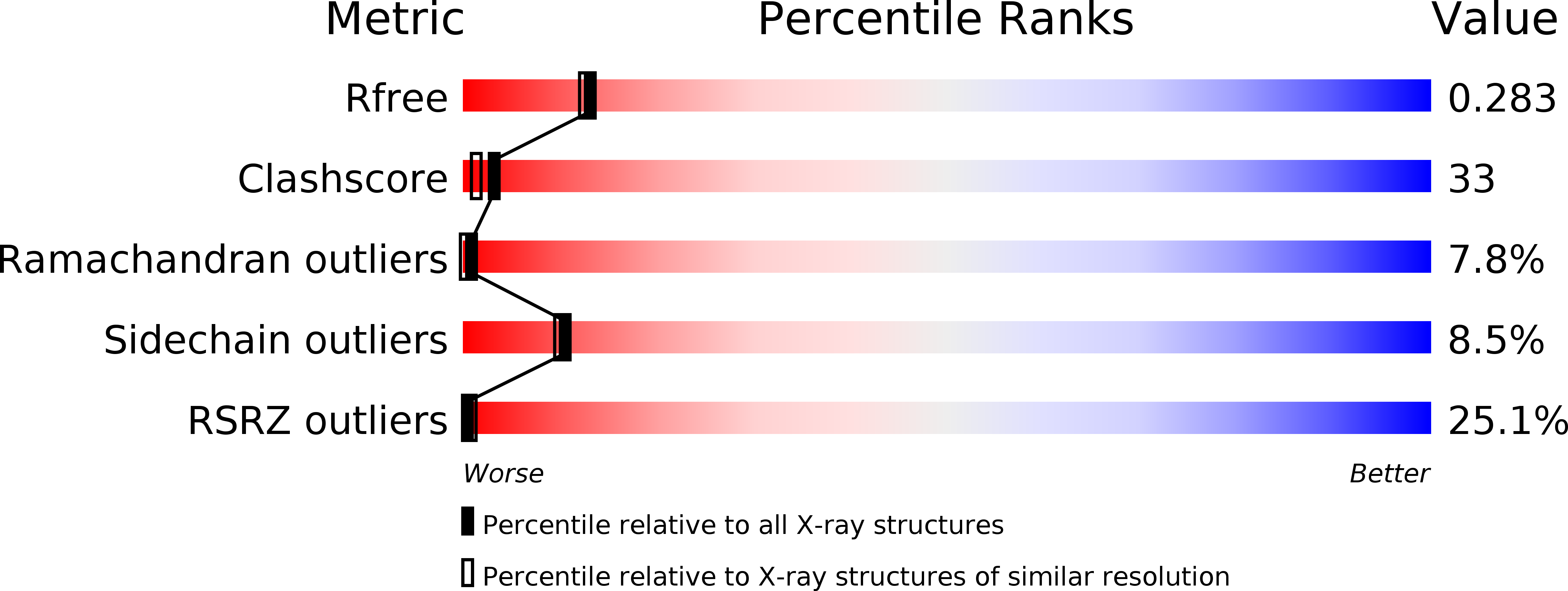
R-Value Free:
0.31
R-Value Work:
0.25
R-Value Observed:
0.25
Space Group:
P 63