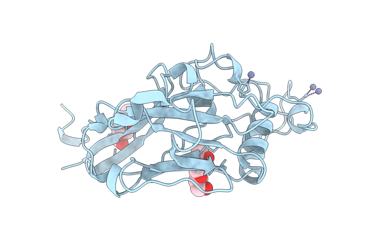
1AOL image
Deposition Date 1997-07-08
Release Date 1997-10-15
Last Version Date 2024-11-06
Entry Detail
PDB ID:
1AOL
Keywords:
Title:
FRIEND MURINE LEUKEMIA VIRUS RECEPTOR-BINDING DOMAIN
Biological Source:
Source Organism:
Host Organism:
Method Details:
Experimental Method:
Resolution:
2.00 Å
R-Value Free:
0.26
R-Value Work:
0.22
R-Value Observed:
0.22
Space Group:
P 21 21 21
Macromolecular Entities
Polymer Type:polypeptide(L)
Molecule:GP70
Gene (Uniprot):env
Chain IDs:A
Chain Length:228
Number of Molecules:1
Biological Source:Friend murine leukemia virus
Modified Residue
Compound ID Chain ID Parent Comp ID Details 2D Image
ASN A ASN GLYCOSYLATION SITE
Primary Citation
Structure of a murine leukemia virus receptor-binding glycoprotein at 2.0 angstrom resolution.
Science 277 1662 1666 (1997)
PMID: 9287219 DOI: 10.1126/science.277.5332.1662

Abstact

An essential step in retrovirus infection is the binding of the virus to its receptor on a target cell. The structure of the receptor-binding domain of the envelope glycoprotein from Friend murine leukemia virus was determined to 2.0 angstrom resolution by x-ray crystallography. The core of the domain is an antiparallel beta sandwich, with two interstrand loops forming a helical subdomain atop the sandwich. The residues in the helical region, but not in the beta sandwich, are highly variable among mammalian C-type retroviruses with distinct tropisms, indicating that the helical subdomain determines the receptor specificity of the virus.

Legend

Protein

Chemical

Disease

Primary Citation of related structures