1ANU image
Deposition Date 1996-07-19
Release Date 1997-07-23
Last Version Date 2024-02-07
Entry Detail
PDB ID:
1ANU
Keywords:
Title:
COHESIN-2 DOMAIN OF THE CELLULOSOME FROM CLOSTRIDIUM THERMOCELLUM
Biological Source:
Source Organism:
Host Organism:
Method Details:
Experimental Method:
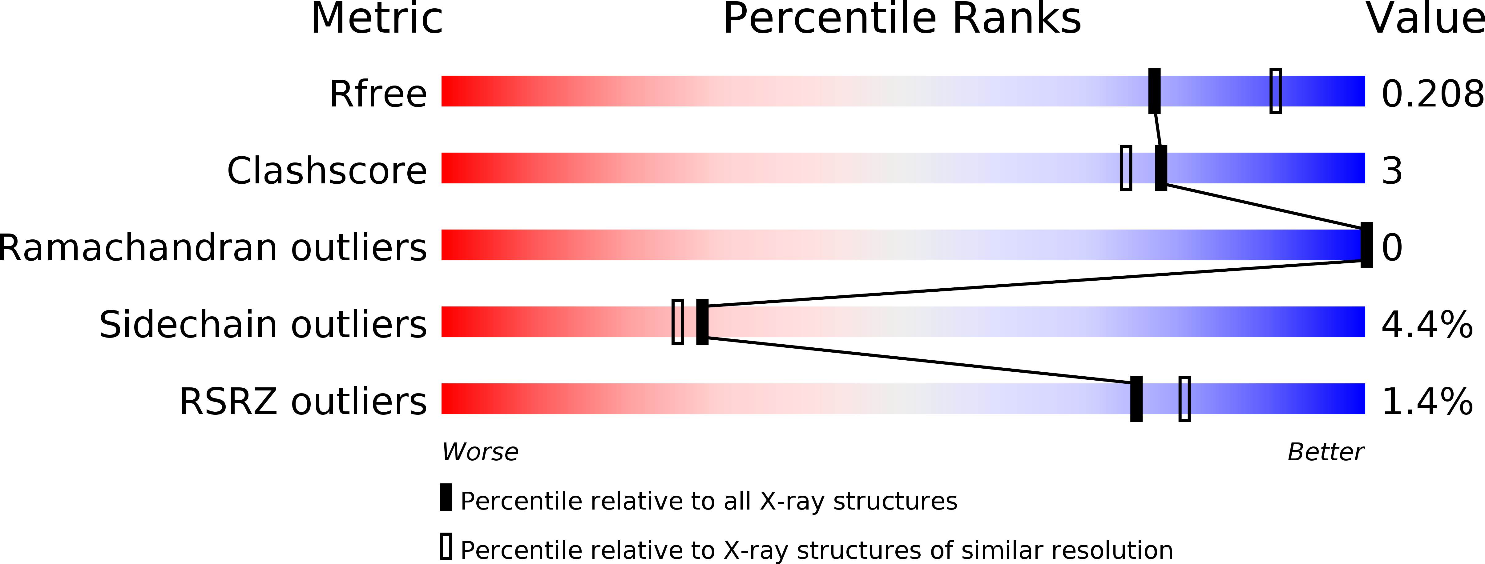
Resolution:
2.15 Å
R-Value Free:
0.21
R-Value Work:
0.19
R-Value Observed:
0.19
Space Group:
C 1 2 1
Macromolecular Entities
Structures with similar UniProt ID
Protein Blast
Polymer Type:polypeptide(L)
Molecule:COHESIN-2
Gene (Uniprot):cipA
Chain IDs:A
Chain Length:138
Number of Molecules:1
Biological Source:Clostridium thermocellum
Primary Citation
A cohesin domain from Clostridium thermocellum: the crystal structure provides new insights into cellulosome assembly.
Structure 5 381 390 (1997)
PMID: 9083107 DOI: 10.1016/S0969-2126(97)00195-0

Abstact

BACKGROUND The scaffoldin component of the cellulolytic bacterium Clostridium thermocellum is a non-hydrolytic protein which organizes the hydrolytic enzymes in a large complex, called the cellulosome. Scaffoldin comprises a series of functional domains, amongst which is a single cellulose-binding domain and nine cohesin domains which are responsible for integrating the individual enzymatic subunits into the complex. The cohesin domains are highly conserved in their primary amino acid sequences. These domains interact with a complementary domain, termed the dockerin domain, one of which is located on each enzymatic subunit. The cohesin-dockerin interaction is the crucial interaction for complex formation in the cellulosome. The determination of structural information about the cohesin domain will provide insights into cellulosome assembly and activity. RESULTS We have determined the three-dimensional crystal structure of one of the cohesin domains from C. thermocellum (cohesin 2) at 2.15 A resolution. The domain forms a nine-stranded beta sandwich with a jelly-roll topology, somewhat similar to the fold displayed by its neighboring cellulose-binding domain. CONCLUSIONS The compact nature of the cohesin structure and its lack of a defined binding pocket suggests that binding between the cohesin and dockerin domains is characterized by interactions between exposed surface residues. As the cohesin-dockerin interaction appears to be rather nonselective, the binding face would presumably be characterized by surface residues which exhibit both intraspecies conservation and interspecies dissimilarity. Within the same species, unconserved surface residues may reflect the position of a given cohesin domain within the scaffoldin subunit, its orientation and interactions with neighboring domains.

Legend

Protein

Chemical

Disease

Primary Citation of related structures
Feedback Form
Name
Email
Institute
Feedback