1AN8 image
Deposition Date 1997-06-27
Release Date 1998-04-29
Last Version Date 2024-02-07
Entry Detail
PDB ID:
1AN8
Title:
CRYSTAL STRUCTURE OF THE STREPTOCOCCAL SUPERANTIGEN SPE-C
Biological Source:
Source Organism:
Host Organism:
Method Details:
Experimental Method:
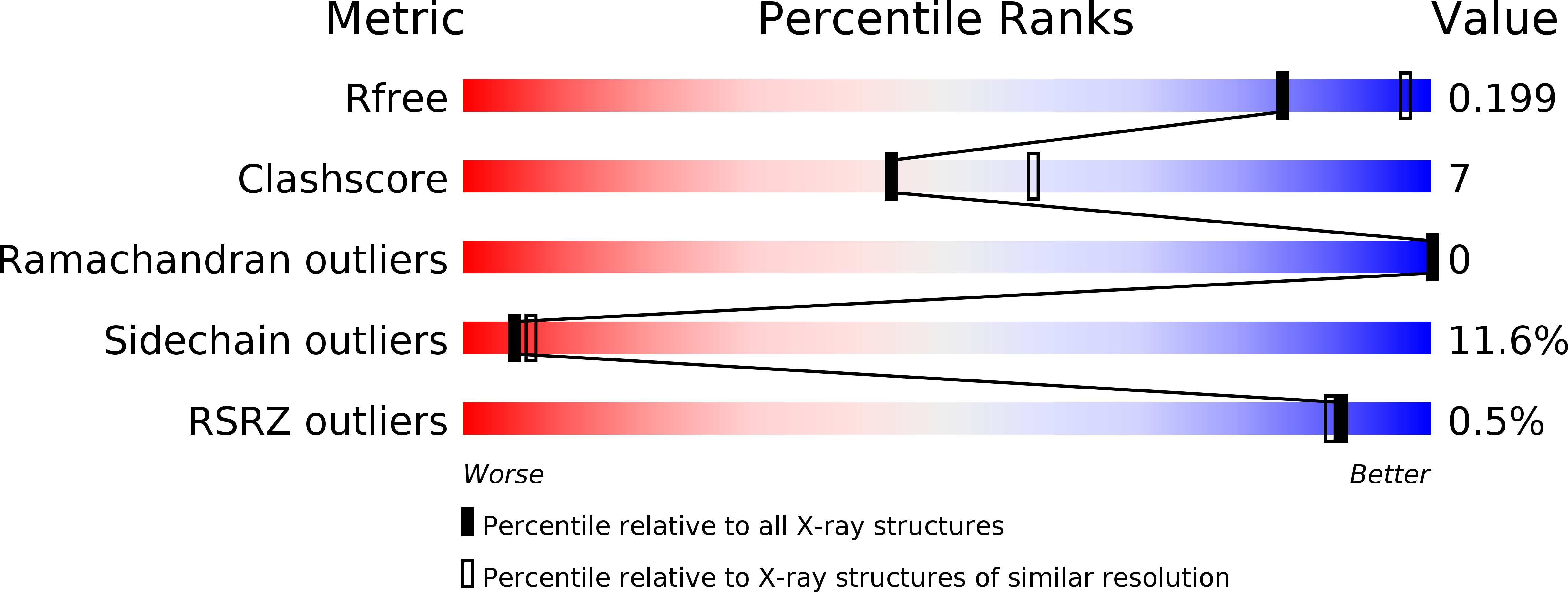
Resolution:
2.40 Å
R-Value Free:
0.21
R-Value Work:
0.17
R-Value Observed:
0.17
Space Group:
P 43 21 2
Macromolecular Entities
Structures with similar UniProt ID
Protein Blast
Polymer Type:polypeptide(L)
Molecule:STREPTOCOCCAL PYROGENIC EXOTOXIN C
Chain IDs:A
Chain Length:208
Number of Molecules:1
Biological Source:Streptococcus pyogenes
Primary Citation
Crystal structure of the streptococcal superantigen SPE-C: dimerization and zinc binding suggest a novel mode of interaction with MHC class II molecules.
Nat.Struct.Biol. 4 635 643 (1997)
PMID: 9253413 DOI: 10.1038/nsb0897-635

Abstact

Bacterial superantigens are small proteins that have a very potent stimulatory effect on T lymphocytes through their ability to bind to both MHC class II molecules and T-cell receptors. We have determined the three-dimensional structure of a Streptococcal superantigen, SPE-C, at 2.4 A resolution. The structure shows that SPE-C has the usual superantigen fold, but that the surface that forms a generic, low-affinity MHC-binding site in other superantigens is here used to create a SPE-C dimer. Instead, MHC class II binding occurs through a zinc binding site that is analogous to a similar site in staphylococcal enterotoxin A. Consideration of the SPE-C dimer suggests a novel mechanism for promotion of MHC aggregation and T-cell activation.

Legend

Protein

Chemical

Disease

Primary Citation of related structures