1AN1 image
Deposition Date 1997-06-26
Release Date 1998-07-01
Last Version Date 2024-11-20
Entry Detail
PDB ID:
1AN1
Title:
LEECH-DERIVED TRYPTASE INHIBITOR/TRYPSIN COMPLEX
Biological Source:
Source Organism:
Sus scrofa (Taxon ID: 9823)
Hirudo medicinalis (Taxon ID: 6421)
Method Details:
Experimental Method:
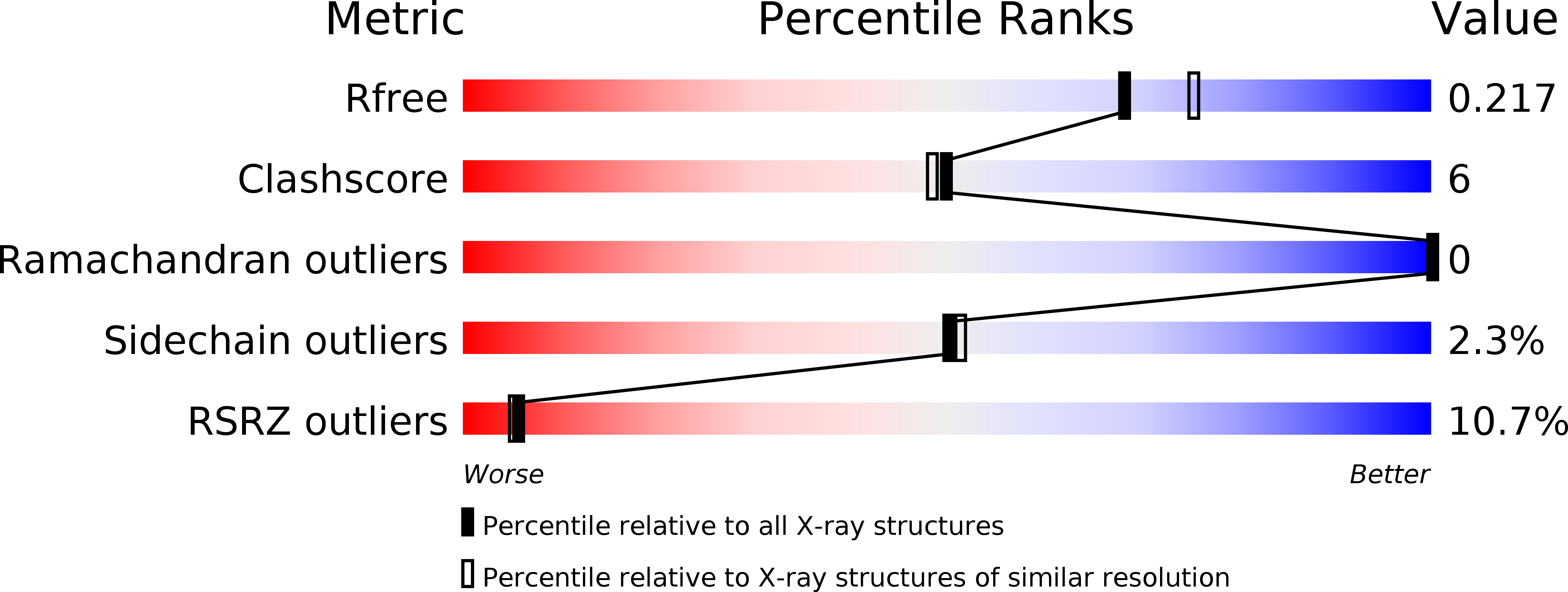
Resolution:
2.03 Å
R-Value Free:
0.22
R-Value Work:
0.17
R-Value Observed:
0.17
Space Group:
P 43 21 2
Macromolecular Entities
Structures with similar UniProt ID
Protein Blast
Polymer Type:polypeptide(L)
Molecule:TRYPSIN
Chain IDs:A (auth: E)
Chain Length:223
Number of Molecules:1
Biological Source:Sus scrofa
Structures with similar UniProt ID
Protein Blast
Polymer Type:polypeptide(L)
Molecule:TRYPTASE INHIBITOR
Mutagens:N115D (D-ASPARTIC ACID FORM)
Chain IDs:B (auth: I)
Chain Length:46
Number of Molecules:1
Biological Source:Hirudo medicinalis
Modified Residue
Compound ID Chain ID Parent Comp ID Details 2D Image
DAS A ASP D-ASPARTIC ACID
Ligand Molecules
Primary Citation
Structure of the complex of leech-derived tryptase inhibitor (LDTI) with trypsin and modeling of the LDTI-tryptase system.
Structure 5 1465 1474 (1997)
PMID: 9384562 DOI: 10.1016/S0969-2126(97)00296-7

Abstact

BACKGROUND Tryptase is a trypsin-like serine proteinase stored in the cytoplasmic granules of mast cells, which has been implicated in a number of mast cell related disorders such as asthma and rheumatoid arthritis. Unlike almost all other serine proteinases, tryptase is fully active in plasma and in the extracellular space, as there are no known natural inhibitors of tryptase in humans. Leech-derived tryptase inhibitor (LDTI), a protein of 46 amino acids, is the first molecule found to bind tightly to and specifically inhibit human tryptase in the nanomolar range. LDTI also inhibits trypsin and chymotrypsin with similar affinities. The structure of LDTI in complex with an inhibited proteinase could be used as a template for the development of low molecular weight tryptase inhibitors. RESULTS The crystal structure of the complex between trypsin and LDTI was solved at 2.0 A resolution and a model of the LDTI-tryptase complex was created, based on this X-ray structure. LDTI has a very similar fold to the third domain of the turkey ovomucoid inhibitor. LDTI interacts with trypsin almost exclusively through its binding loop (residues 3-10) and especially through the sidechain of the specificity residue Lys8. Our modeling studies indicate that these interactions are maintained in the LDTI-tryptase complex. CONCLUSIONS The insertion of nine residues after residue 174 in tryptase, relative to trypsin and chymotrypsin, prevents inhibition by other trypsin inhibitors and is certainly responsible for the higher specificity of tryptase relative to trypsin. In LDTI, the disulfide bond between residues 4 and 25 causes a sharp turn from the binding loop towards the N terminus, holding the N terminus away from the 174 loop of tryptase.

Legend

Protein

Chemical

Disease

Primary Citation of related structures
Feedback Form
Name
Email
Institute
Feedback