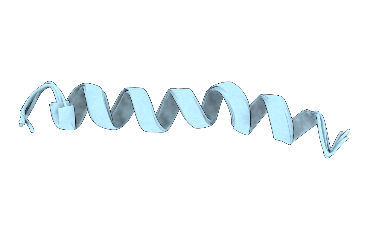
1AMC image
Deposition Date 1994-11-14
Release Date 1995-01-26
Last Version Date 2024-05-22
Entry Detail
PDB ID:
1AMC
Title:
SOLUTION STRUCTURE OF RESIDUES 1-28 OF THE AMYLOID BETA-PEPTIDE
Biological Source:
Source Organism:
Homo sapiens (Taxon ID: 9606)
Method Details:
Experimental Method:
Conformers Submitted:
5
Macromolecular Entities
Polymer Type:polypeptide(L)
Molecule:AMYLOID BETA-PEPTIDE
Gene (Uniprot):APP
Chain IDs:A
Chain Length:28
Number of Molecules:1
Biological Source:Homo sapiens
Ligand Molecules
Primary Citation
Solution structure of residues 1-28 of the amyloid beta-peptide.
Biochemistry 33 7788 7796 (1994)
PMID: 7516706 DOI: 10.1021/bi00191a006

Abstact

The three-dimensional solution structure of residues 1-28 of the amyloid beta-peptide was determined using nuclear magnetic resonance spectroscopy, distance geometry, and molecular dynamic techniques. The nuclear magnetic resonance data used to derive the structure consisted of nuclear Overhauser enhancements, vicinal coupling constants, and temperature coefficients of the amide-NH chemical shifts. The beta-peptide is the major proteinaceous component of amyloid deposits in Alzheimer's disease. In membrane-like media, the peptide folds to form a predominately alpha-helical structure with a bend centered at residue 12. The side chains of histidine-13 and lysine-16 are close, residing on the same face of the helix. Their proximity may constitute a binding motif with the heparan sulfate proteoglycans. The molecular details of the structure shown here could facilitate the design of rational treatments to curtail the binding of heparan sulfate proteoglycans or to prevent an alpha-helix-->beta-sheet conversion that may occur during the early stages of amyloid formation in Alzheimer's disease.

Legend

Protein

Chemical

Disease

Primary Citation of related structures