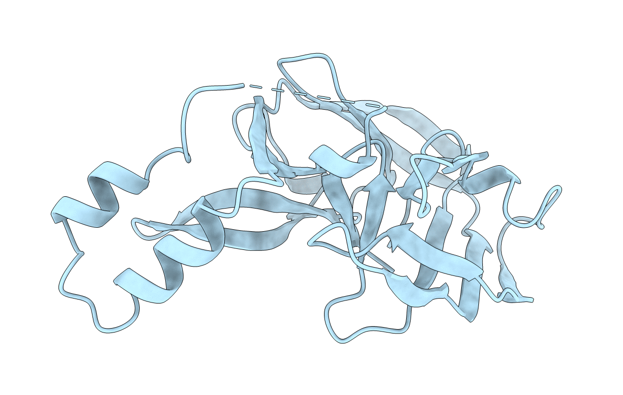
1AM2 image
Deposition Date 1997-06-20
Release Date 1998-06-24
Last Version Date 2024-02-07
Entry Detail
PDB ID:
1AM2
Keywords:
Title:
GYRA INTEIN FROM MYCOBACTERIUM XENOPI
Biological Source:
Source Organism:
Host Organism:
Method Details:
Experimental Method:
Resolution:
2.20 Å
R-Value Free:
0.25
R-Value Work:
0.18
R-Value Observed:
0.18
Space Group:
P 32 2 1
Macromolecular Entities
Polymer Type:polypeptide(L)
Molecule:MXE GYRA INTEIN
Gene (Uniprot):gyrA
Mutations:Y0A, C1S
Chain IDs:A
Chain Length:199
Number of Molecules:1
Biological Source:Mycobacterium xenopi
Primary Citation
Crystal structure of GyrA intein from Mycobacterium xenopi reveals structural basis of protein splicing.
Nat.Struct.Biol. 5 31 36 (1998)
PMID: 9437427 DOI: 10.1038/nsb0198-31

Abstact

Several genes from prokaryotes and lower eukaryotes have been found to contain an in-frame open reading frame, which encodes for an internal protein (intein). Post-translationally, the internal polypeptide auto-splices and ligates the external sequences to yield a functional external protein (extein) and an intein. Most, but not all inteins, contain, apart from a splicing domain, a separate endonucleolytic domain that enables them to maintain their presence by a homing mechanism. We report here the crystal structure of an intein found in the gyrase A subunit from Mycobacterium xenopi at 2.2 A resolution. The structure contains an unusual beta-fold with the catalytic splice junctions at the ends of two adjacent antiparallel beta-strands. The arrangement of the active site residues Ser 1, Thr 72, His 75, His 197, and Asn 198 is consistent with a four-step mechanism for the cleavage-ligation reaction. Using site-directed mutagenesis, the N-terminal cysteine, proposed as the nucleophile in the first step of the splicing reaction, was changed to a Ser 1 and Ala 0, thus capturing the intein in a pre-spliced state.

Legend

Protein

Chemical

Disease

Primary Citation of related structures