Deposition Date
1997-06-10
Release Date
1997-12-24
Last Version Date
2024-02-07
Entry Detail
PDB ID:
1AL3
Keywords:
Title:
COFACTOR BINDING FRAGMENT OF CYSB FROM KLEBSIELLA AEROGENES
Biological Source:
Source Organism(s):
Klebsiella aerogenes (Taxon ID: 28451)
Expression System(s):
Method Details:
Experimental Method:
Resolution:
1.80 Å
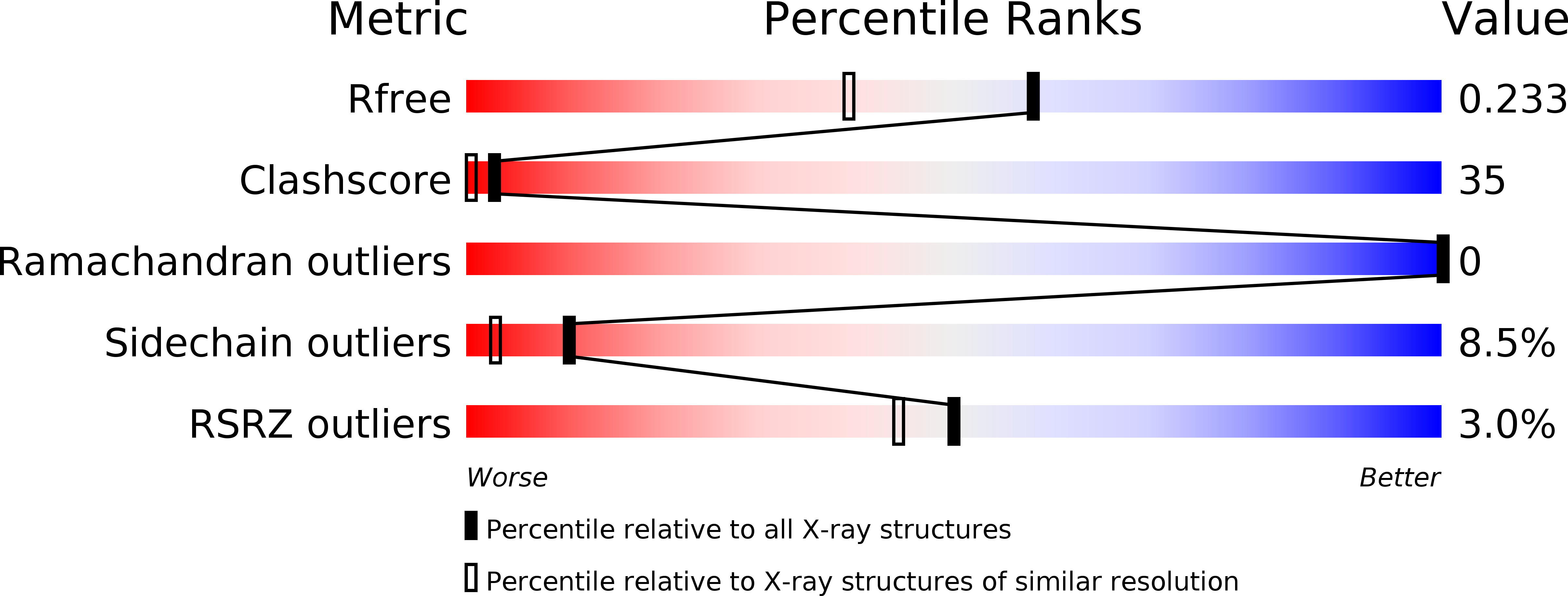
R-Value Free:
0.24
R-Value Work:
0.17
Space Group:
P 21 21 2