1AKG image
Deposition Date 1997-05-18
Release Date 1998-05-20
Last Version Date 2024-10-23
Entry Detail
PDB ID:
1AKG
Title:
ALPHA-CONOTOXIN PNIB FROM CONUS PENNACEUS
Biological Source:
Source Organism:
Method Details:
Experimental Method:
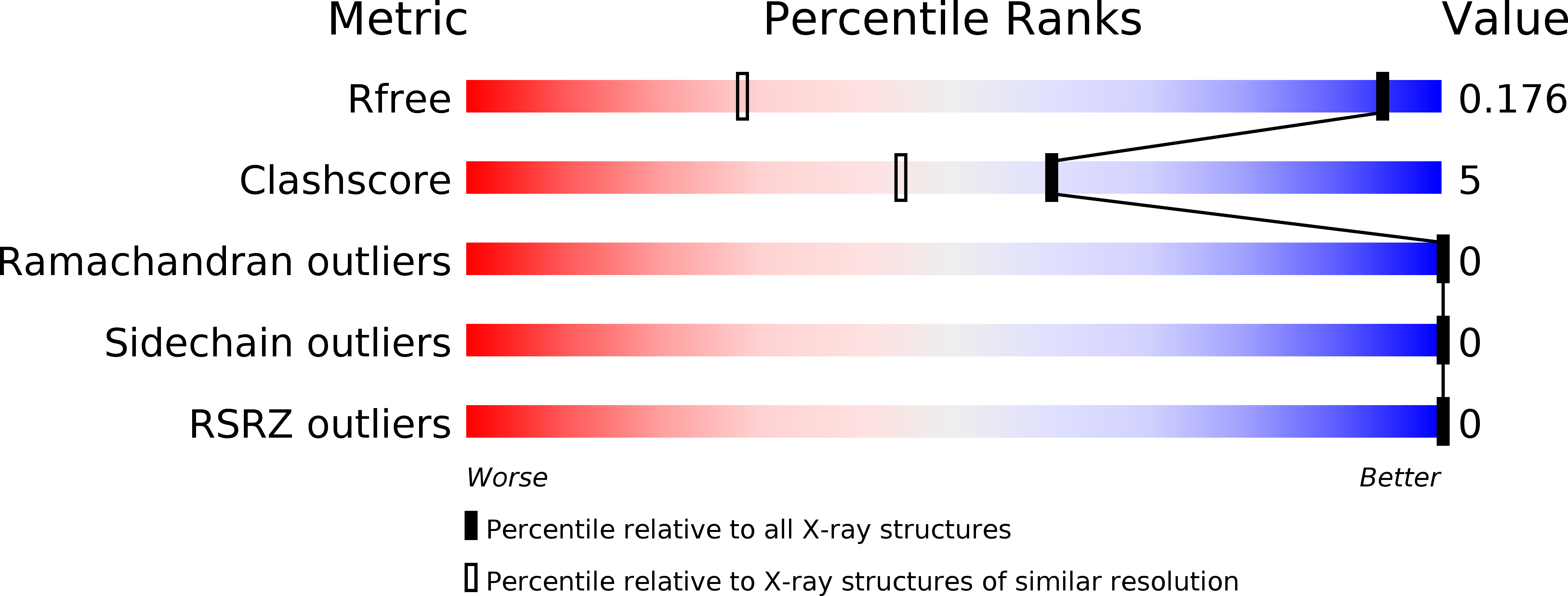
Resolution:
1.10 Å
R-Value Free:
0.15
R-Value Work:
0.14
R-Value Observed:
0.14
Space Group:
P 21 21 21
Macromolecular Entities
Structures with similar UniProt ID
Protein Blast
Polymer Type:polypeptide(L)
Molecule:ALPHA-CONOTOXIN PNIB
Chain IDs:A
Chain Length:17
Number of Molecules:1
Biological Source:Conus pennaceus
Primary Citation
Crystal structure at 1.1 A resolution of alpha-conotoxin PnIB: comparison with alpha-conotoxins PnIA and GI.
Biochemistry 36 11323 11330 (1997)
PMID: 9298951 DOI: 10.1021/bi9713052

Abstact

Conotoxins are small, cysteine-rich peptides isolated from the venom of Conus spp. of predatory marine snails, which selectively target specific receptors and ion channels critical to the functioning of the neuromuscular system. alpha-Conotoxins PnIA and PnIB are both 16-residue peptides (differing in sequence at only two positions) isolated from the molluscivorous snail Conus pennaceus. In contrast to the muscle-selective alpha-conotoxin GI from Conus geographus, PnIA and PnIB block the neuronal nicotinic acetylcholine receptor (nAChR). Here, we describe the crystal structure of PnIB, solved at a resolution of 1.1 A and phased using the Shake-and-Bake direct methods program. PnIB crystals are orthorhombic and belong to the space group P212121 with the following unit cell dimensions: a = 14.6 A, b = 26.1 A, and c = 29.2 A. The final refined structure of alpha-conotoxin PnIB includes all 16 residues plus 23 solvent molecules and has an overall R-factor of 14.7% (R-free of 15.9%). The crystal structures of the alpha-conotoxins PnIB and PnIA are solved from different crystal forms, with different solvent contents. Comparison of the structures reveals them to be very similar, showing that the unique backbone and disulfide architecture is not strongly influenced by crystal lattice constraints or solvent interactions. This finding supports the notion that this structural scaffold is a rigid support for the presentation of important functional groups. The structures of PnIB and PnIA differ in their shape and surface charge distribution from that of GI.

Legend

Protein

Chemical

Disease

Primary Citation of related structures
Feedback Form
Name
Email
Institute
Feedback