Deposition Date
1991-11-08
Release Date
1994-01-31
Last Version Date
2024-12-25
Entry Detail
PDB ID:
1AKE
Keywords:
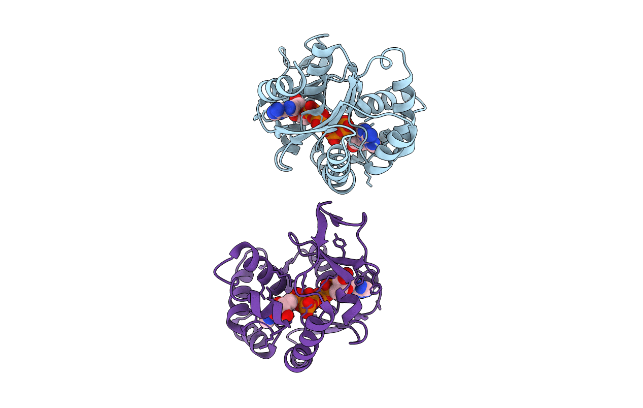
Title:
STRUCTURE OF THE COMPLEX BETWEEN ADENYLATE KINASE FROM ESCHERICHIA COLI AND THE INHIBITOR AP5A REFINED AT 1.9 ANGSTROMS RESOLUTION: A MODEL FOR A CATALYTIC TRANSITION STATE
Biological Source:
Source Organism(s):
Escherichia coli (Taxon ID: 562)
Expression System(s):
Method Details:
Experimental Method:
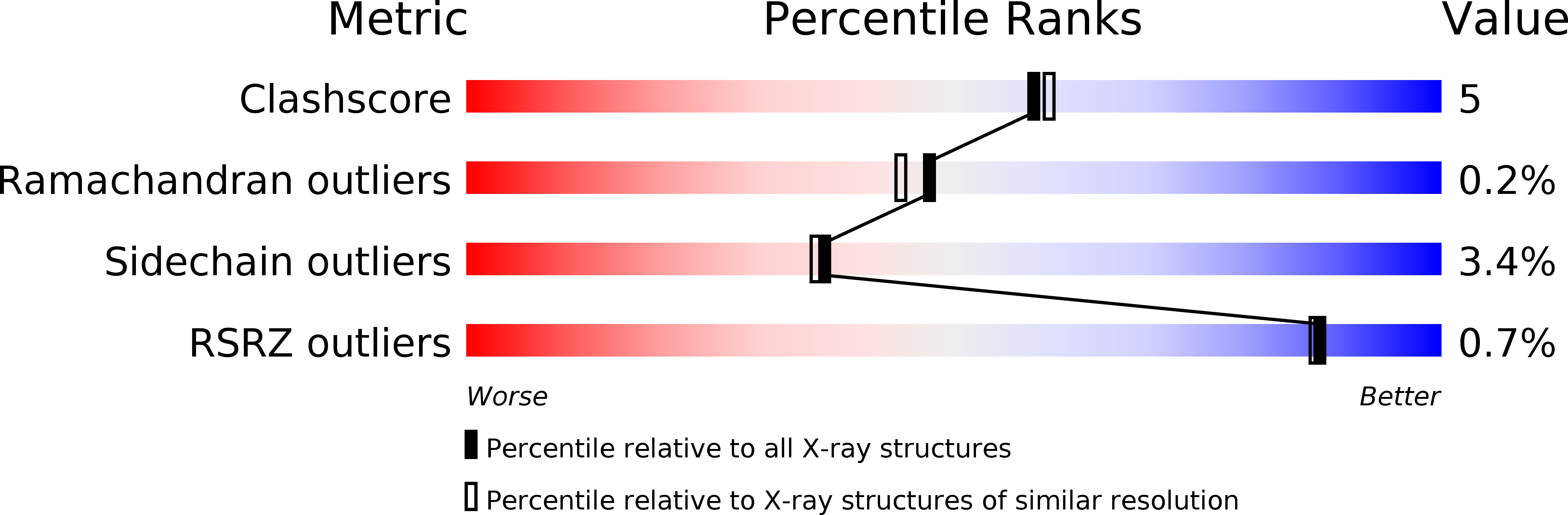
Resolution:
2.00 Å
R-Value Work:
0.19
R-Value Observed:
0.19
Space Group:
P 21 2 21