1AKD image
Deposition Date 1997-05-16
Release Date 1997-11-19
Last Version Date 2024-04-03
Entry Detail
PDB ID:
1AKD
Keywords:
Title:
CYTOCHROME P450CAM FROM PSEUDOMONAS PUTIDA, COMPLEXED WITH 1S-CAMPHOR
Biological Source:
Source Organism:
Host Organism:
Method Details:
Experimental Method:
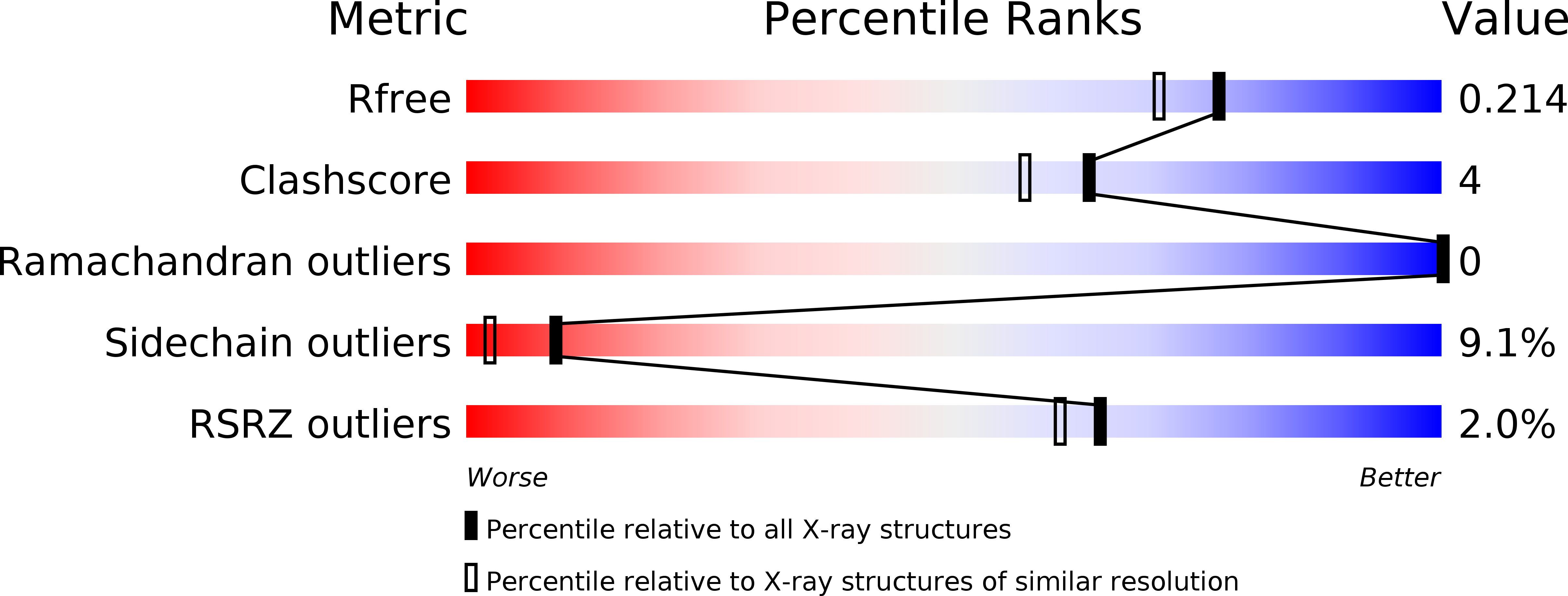
Resolution:
1.80 Å
R-Value Free:
0.26
R-Value Work:
0.20
R-Value Observed:
0.20
Space Group:
P 21 21 21
Macromolecular Entities
Structures with similar UniProt ID
Protein Blast
Polymer Type:polypeptide(L)
Molecule:CYTOCHROME P450CAM
Gene (Uniprot):camC
Chain IDs:A
Chain Length:414
Number of Molecules:1
Biological Source:Pseudomonas putida
Primary Citation
Crystal structure of cytochrome P-450cam complexed with the (1S)-camphor enantiomer.
FEBS Lett. 415 253 257 (1997)
PMID: 9357977 DOI: 10.1016/S0014-5793(97)01135-6

Abstact

The crystal structure of cytochrome P-450cam complexed with the enantiomer (1S)-camphor has been solved to 1.8 angstroms resolution and compared with the structure of the (1R)-camphor P-450cam complex. The overall protein structure is the same for both enantiomer complexes. However, the orientation of the substrates in the heme pocket differs. In contrast to (1R)-camphor, the (1S)-enantiomer binds in at least two orientations. The major binding mode of (1S)-camphor resembles the one of the (1R)-enantiomer in that there is a hydrogen bond between Tyr-96 and the quinone group of camphor, and the 10-methyl group points towards the I-helix. The binding differs in that C-5 is not at a position suitable for hydroxylation. In the other orientation (1S)-camphor is not hydrogen bonded, but C-5 is located suitably for hydroxylation.

Legend

Protein

Chemical

Disease

Primary Citation of related structures