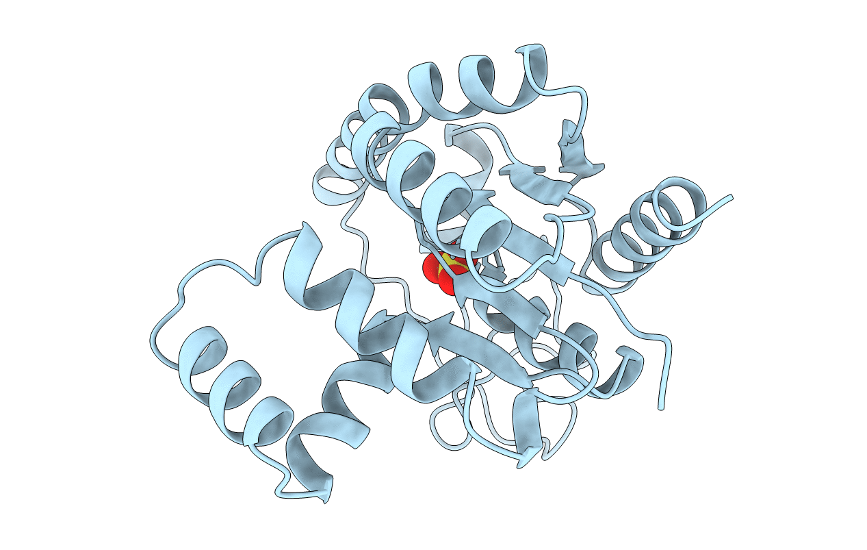
1AK2 image
Deposition Date 1995-12-29
Release Date 1996-06-10
Last Version Date 2024-10-30
Entry Detail
PDB ID:
1AK2
Title:
ADENYLATE KINASE ISOENZYME-2
Biological Source:
Source Organism:
Bos taurus (Taxon ID: 9913)
Method Details:
Experimental Method:
Resolution:
1.92 Å
R-Value Work:
0.22
R-Value Observed:
0.22
Space Group:
P 21 21 21
Macromolecular Entities
Structures with similar UniProt ID
Protein Blast
Polymer Type:polypeptide(L)
Molecule:ADENYLATE KINASE ISOENZYME-2
Gene (Uniprot):AK2
Chain IDs:A
Chain Length:233
Number of Molecules:1
Biological Source:Bos taurus
Ligand Molecules
Primary Citation
The structure of bovine mitochondrial adenylate kinase: comparison with isoenzymes in other compartments.
Protein Sci. 5 434 441 (1996)
PMID: 8868479

Abstact

In vertebrates, there are different adenylate kinases in the compartments cytosol, mitochondrial intermembrane space, and mitochondrial matrix. Here, we report the spatial structure of the intermembrane species established in two crystal forms by X-ray diffraction analyses at 1.92 and 2.1 A resolution. In both structures, the enzyme is unligated, and thus in an "open" conformation. The enzyme was prepared from bovine liver, containing at least five variants arisen from posttranscriptional and posttranslational modifications. It could only be crystallized after removing some of these variants. A comparison with the known structures of the adenylate kinases from cytosol and mitochondrial matrix reveals structural differences that should play a role in protein targeting because none of these enzymes contains a cleavable signal peptide. A further comparison with adenylate kinases from Gram-positive bacteria showed that the structural Zn2+ ion of these species is replaced by a strictly conserved assembly of hydrogen bonded residues.

Legend

Protein

Chemical

Disease

Primary Citation of related structures