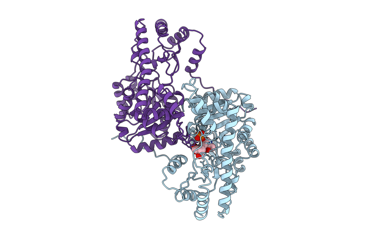
1AJS image
Deposition Date 1997-05-08
Release Date 1997-08-20
Last Version Date 2024-06-05
Entry Detail
PDB ID:
1AJS
Title:
REFINEMENT AND COMPARISON OF THE CRYSTAL STRUCTURES OF PIG CYTOSOLIC ASPARTATE AMINOTRANSFERASE AND ITS COMPLEX WITH 2-METHYLASPARTATE
Biological Source:
Source Organism:
Sus scrofa (Taxon ID: 9823)
Method Details:
Experimental Method:
Resolution:
1.60 Å
R-Value Observed:
0.17
Space Group:
P 21 21 21
Macromolecular Entities
Polymer Type:polypeptide(L)
Molecule:ASPARTATE AMINOTRANSFERASE
Gene (Uniprot):GOT1
Chain IDs:A
Chain Length:412
Number of Molecules:1
Biological Source:Sus scrofa
Polymer Type:polypeptide(L)
Molecule:ASPARTATE AMINOTRANSFERASE
Gene (Uniprot):GOT1
Chain IDs:B
Chain Length:412
Number of Molecules:1
Biological Source:Sus scrofa
Modified Residue
Compound ID Chain ID Parent Comp ID Details 2D Image
LLP B LYS ?
Ligand Molecules
Primary Citation
Refinement and comparisons of the crystal structures of pig cytosolic aspartate aminotransferase and its complex with 2-methylaspartate.
J.Biol.Chem. 272 17293 17302 (1997)
PMID: 9211866 DOI: 10.1074/jbc.272.28.17293

Abstact

Two high resolution crystal structures of cytosolic aspartate aminotransferase from pig heart provide additional insights into the stereochemical mechanism for ligand-induced conformational changes in this enzyme. Structures of the homodimeric native structure and its complex with the substrate analog 2-methylaspartate have been refined, respectively, with 1.74-A x-ray diffraction data to an R value of 0.170, and with 1.6-A data to an R value of 0.173. In the presence of 2-methylaspartate, one of the subunits (subunit 1) shows a ligand-induced conformational change that involves a large movement of the small domain (residues 12-49 and 327-412) to produce a "closed" conformation. No such transition is observed in the other subunit (subunit 2), because crystal lattice contacts lock it in an "open" conformation like that adopted by subunit 1 in the absence of substrate. By comparing the open and closed forms of cAspAT, we propose a stereochemical mechanism for the open-to-closed transition that involves the electrostatic neutralization of two active site arginine residues by the negative charges of the incoming substrate, a large change in the backbone (phi,psi) conformational angles of two key glycine residues, and the entropy-driven burial of a stretch of hydrophobic residues on the N-terminal helix. The calculated free energy for the burial of this "hydrophobic plug" appears to be sufficient to serve as the driving force for domain closure.

Legend

Protein

Chemical

Disease

Primary Citation of related structures