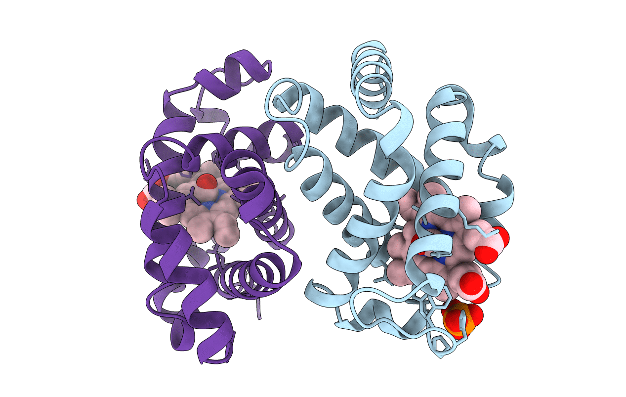
The three-dimensional structure and associated solvent of human carboxyhemoglobin at 2.2 A resolution are compared with other R-state and T-state human hemoglobin structures. The crystal form is isomorphous with that of the 2.7 A structure of carboxyhemoglobin reported earlier [Baldwin (1980). J. Mol. Biol. 136, 103-128], whose coordinates were used as a starting model, and with the 2.2 A structure described in an earlier report [Derewenda et al. (1990). J. Mol. Biol. 211, 515-519]. During the course of the refinement, a natural mutation of the alpha-subunit, A53S, was discovered that forms a new crystal contact through a bridging water molecule. The protein structure shows a significant difference between the alpha and beta heme geometries, with Fe-C-O angles of 125 and 162 degrees, respectively. The carboxyhemoglobin is compared with other fully ligated R-state human hemoglobins [Baldwin (1980). J. Mol. Biol. 136, 103-128; Shaanan (1983). J. Mol. Biol. 195, 419-422] with the R2-state hemoglobin [Silva et al. (1992). J. Biol. Chem. 267, 17248-17256] and with T-state deoxyhemoglobin [Fronticelli et al. (1994). J. Biol. Chem. 269, 23965-23969]. The structure is similar to the earlier reported R-state structures, but there are differences in many side-chain conformations, the associated water structure and the presence and the position of a phosphate ion. The quaternary changes between the R-state carboxyhemoglobin and the R2-state and T-state structures are in general consistent with those reported in the earlier structures. The location of 238 water molecules and a phosphate ion in the carboxyhemoglobin structure allows the first comparison of the solvent structures of the R-state and T-state structures. Distinctive hydration patterns for each of the quaternary structures are observed, but a number of conserved water molecule binding sites are found that are independent of the conformational state of the protein.
Legend
Protein
Chemical
Disease