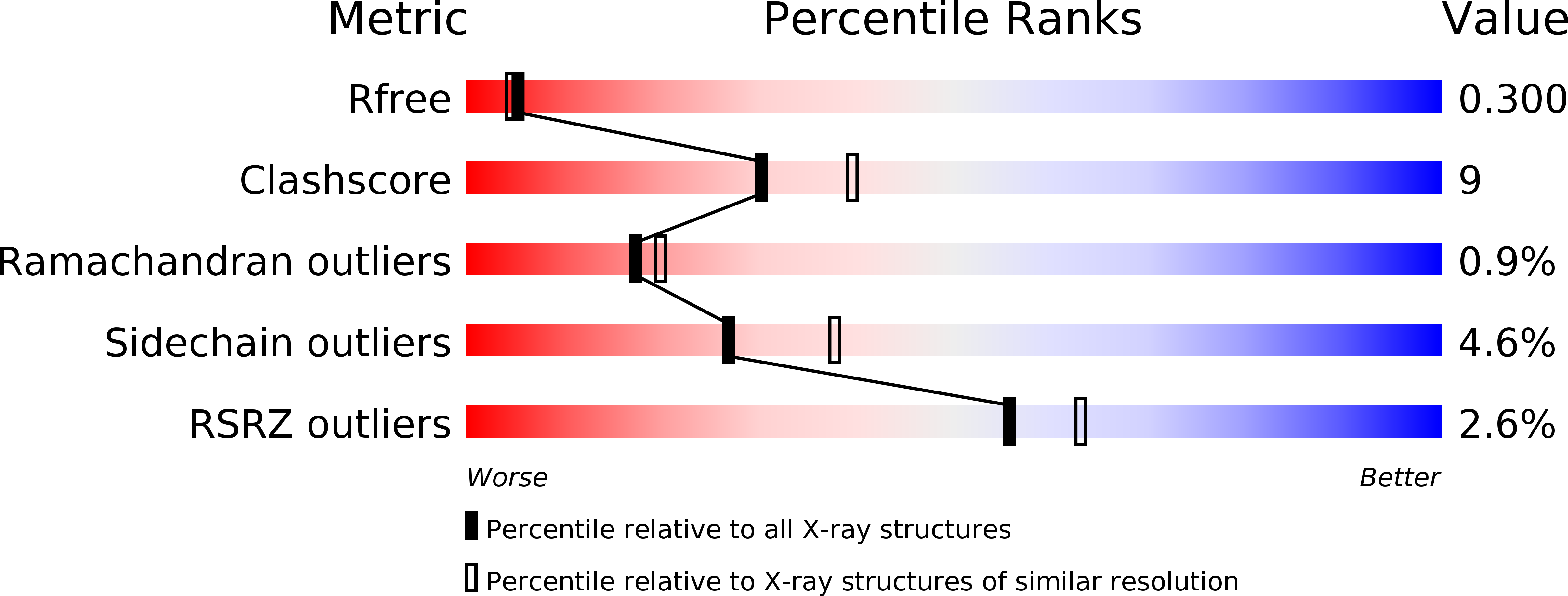
1AJ5 image
Deposition Date 1997-05-15
Release Date 1998-05-20
Last Version Date 2024-02-07
Entry Detail
PDB ID:
1AJ5
Title:
CALPAIN DOMAIN VI APO
Biological Source:
Source Organism:
Method Details:
Experimental Method:
Resolution:
2.30 Å
R-Value Free:
0.30
R-Value Work:
0.22
R-Value Observed:
0.22
Space Group:
C 2 2 21
Macromolecular Entities
Structures with similar UniProt ID
Protein Blast
Polymer Type:polypeptide(L)
Molecule:CALPAIN
Gene (Uniprot):Capns1
Chain IDs:A, B
Chain Length:173
Number of Molecules:2
Biological Source:Rattus norvegicus
Primary Citation
Structure of a calpain Ca(2+)-binding domain reveals a novel EF-hand and Ca(2+)-induced conformational changes.
Nat.Struct.Biol. 4 532 538 (1997)
PMID: 9228945 DOI: 10.1038/nsb0797-532

Abstact

The crystal structure of a Ca(2+)-binding domain (dVI) of rat m-calpain has been determined at 2.3 A resolution, both with and without bound Ca2+. The structures reveal a unique fold incorporating five EF-hand motifs per monomer, three of which bind calcium at physiological calcium concentrations, with one showing a novel EF-hand coordination pattern. This investigation gives us a first view of the calcium-induced conformational changes, and consequently an insight into the mechanism of calcium induced activation in calpain. The crystal structures reveal a dVI homodimer which provides a preliminary model for the subunit dimerization in calpain.

Legend

Protein

Chemical

Disease

Primary Citation of related structures
Feedback Form
Name
Email
Institute
Feedback