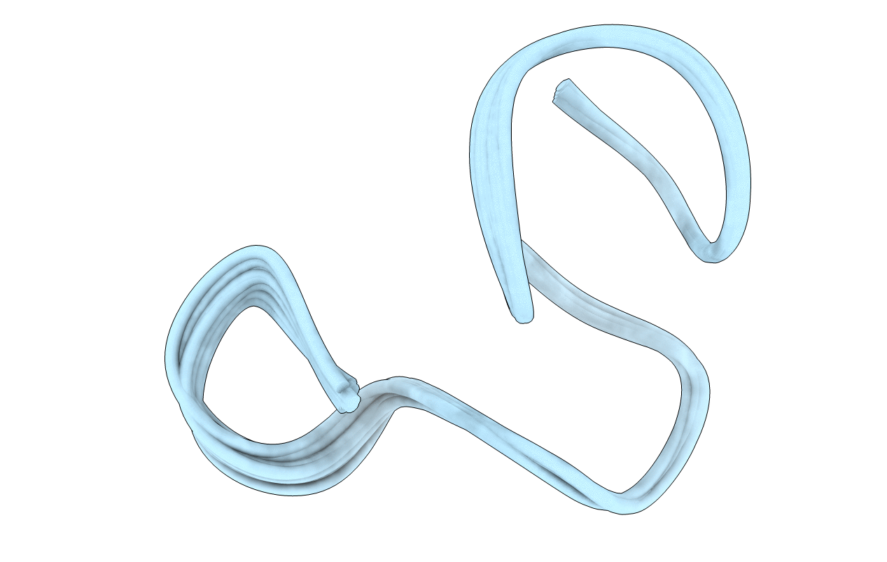
1AJ1 image
Entry Detail
PDB ID:
1AJ1
Keywords:
Title:
NMR STRUCTURE OF THE LANTIBIOTIC ACTAGARDINE
Biological Source:
Source Organism:
PDB Version:
Deposition Date:
1997-05-14
Release Date:
1997-10-15
Method Details:
Experimental Method:
Conformers Calculated:
100
Conformers Submitted:
15
Selection Criteria:
LEAST RESTRAINT VIOLATIONS, BEST RELAXATION MATRIX R- FACTORS
Macromolecular Entities
Polymer Type:polypeptide(L)
Description:LANTIBIOTIC ACTAGARDINE
Chain IDs:A
Chain Length:19
Number of Molecules:1
Biological Source:Actinoplanes liguriensis
Modified Residue
Compound ID Chain ID Parent Comp ID Details 2D Image
DBB A THR POST-TRANSLATIONAL MODIFICATION
Ligand Molecules
Peptide-like Molecules
PRD_000194
Primary Citation
The three-dimensional solution structure of the lantibiotic murein-biosynthesis-inhibitor actagardine determined by NMR.
Eur.J.Biochem. 246 809 819 (1997)
PMID: 9219543 DOI: 10.1111/j.1432-1033.1997.00809.x

Abstact

The three-dimensional solution structure of the lantibiotic actagardine was determined at high resolution by homonuclear and heteronuclear two-dimensional and three-dimensional NMR spectroscopy in [2H3]acetonitrile/H2O (7:3). 133 non-trivial distance and 22 torsional-angle constraints were derived from the NMR data. An ensemble of 15 low-energy structures was calculated by distance geometry followed by an iterative relaxation-matrix-refinement procedure. The rmsd of the backbone coordinates with respect to the average structure was 17 pm. The two distinct thioether ring systems 1-6 and 7-19 were even better defined, with backbone rmsd of 10 pm and 14 pm, respectively. Actagardine shows a rigid compact globular shape based on the constraining bridging pattern, which is composed of an N-terminal lanthionine ring from residues 1-6 and three intertwined C-terminal methyllanthionine rings comprising residues 7-12, 9-17 and 14-19. In addition, this C-terminal ring system is stabilised by a short antiparallel beta sheet. A feature of the actagardine structure is the presence of two putative binding pockets. A pocket is generated by the covalent constraints of the C-terminal thioether ring system. The rim of this pocket is built up by a loop structure comprising residues 12-19, whose backbone amide protons are all directed to the centre of the pocket. The second pocket is formed by an L-shaped orientation of the N-terminal and C-terminal thioether ring systems. The only two hydrophilic amino acid residues of actagardine, Glu11 and Ser2, are directed to this pocket. A region of high sequence similarity with the related lantibiotic mersacidin is located exactly at the position of the second pocket (residues 3-12). This suggests that the second pocket is responsible for the antibiotic mode of action of actagardine and mersacidin as inhibitors of the murein biosynthesis of gram-positive bacteria.

Legend

Protein

Chemical

Disease

Primary Citation of related structures