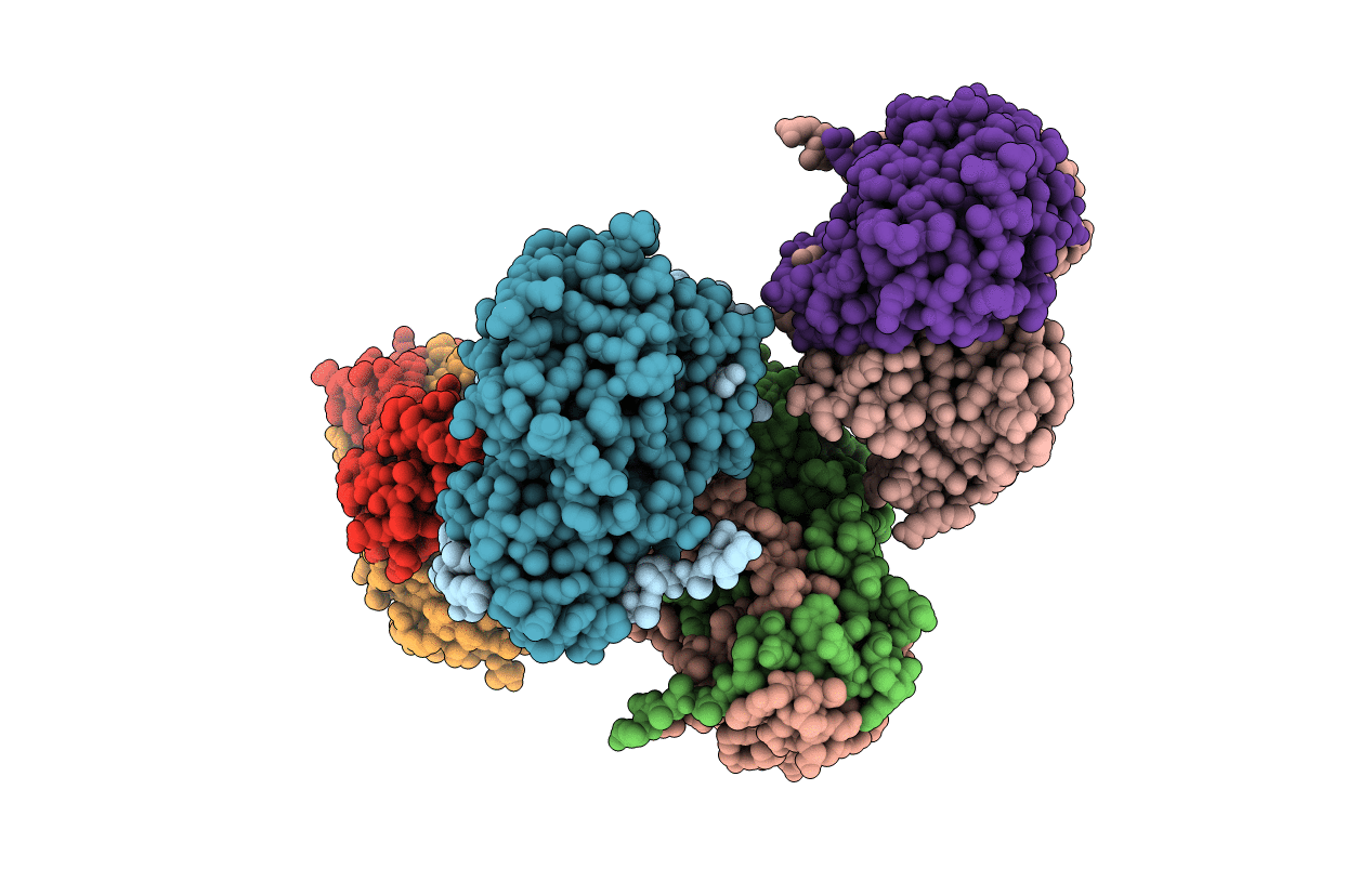
1AHJ image
Deposition Date 1997-04-05
Release Date 1998-04-08
Last Version Date 2024-10-30
Entry Detail
PDB ID:
1AHJ
Keywords:
Title:
NITRILE HYDRATASE
Biological Source:
Source Organism:
Method Details:
Experimental Method:
Resolution:
2.65 Å
R-Value Free:
0.28
R-Value Work:
0.26
R-Value Observed:
0.26
Space Group:
P 21 21 21
Macromolecular Entities
Structures with similar UniProt ID
Protein Blast
Polymer Type:polypeptide(L)
Molecule:NITRILE HYDRATASE (SUBUNIT ALPHA)
Gene (Uniprot):nthA
Chain IDs:A, C, E, G
Chain Length:207
Number of Molecules:4
Biological Source:Rhodococcus sp. R312
Structures with similar UniProt ID
Protein Blast
Polymer Type:polypeptide(L)
Molecule:NITRILE HYDRATASE (SUBUNIT BETA)
Gene (Uniprot):nthB
Chain IDs:B, D, F, H
Chain Length:212
Number of Molecules:4
Biological Source:Rhodococcus sp. R312
Ligand Molecules
Primary Citation
Crystal structure of nitrile hydratase reveals a novel iron centre in a novel fold.
Structure 5 691 699 (1997)
PMID: 9195885 DOI: 10.1016/S0969-2126(97)00223-2

Abstact

BACKGROUND Nitrile hydratases are unusual metalloenzymes that catalyze the hydration of nitriles to their corresponding amides. They are used as biocatalysts in acrylamide production, one of the few commercial scale bioprocesses, as well as in environmental remediation for the removal of nitriles from waste streams. Nitrile hydratases are composed of two subunits, alpha and beta, and they contain one iron atom per alphabeta unit. We have determined the crystal structure of photoactivated iron-containing nitrile hydratase from Rhodococcus sp. R312 to 2.65 A resolution as a first step in the elucidation of its catalytic mechanism. RESULTS The alpha subunit consists of a long N-terminal arm and a C-terminal domain that forms a novel fold. This fold can be described as a four layered structure, alpha-beta-beta-alpha, with unusual connectivities between the beta strands. The beta subunit also contains a long N-terminal extension, a helical domain, and a C-terminal domain that folds into a beta roll. The two subunits form a tight heterodimer that is the functional unit of the enzyme. The active site is located in a cavity at the subunit-subunit interface. The iron centre is formed by residues from the alpha subunit only-three cysteine thiolates and two mainchain amide nitrogen atoms are ligands. CONCLUSIONS Nitrile hydratases contain a novel iron centre with a structure not previously observed in proteins; it resembles a hybrid of the iron centres of heme and Fe-S proteins. The low-spin electronic configuration presumably results in part from two Fe-amide nitrogen bonds. The structure is consistent with the metal ion having a role as a Lewis acid in the catalytic reaction.

Legend

Protein

Chemical

Disease

Primary Citation of related structures