Deposition Date
1997-03-28
Release Date
1997-06-16
Last Version Date
2024-05-22
Entry Detail
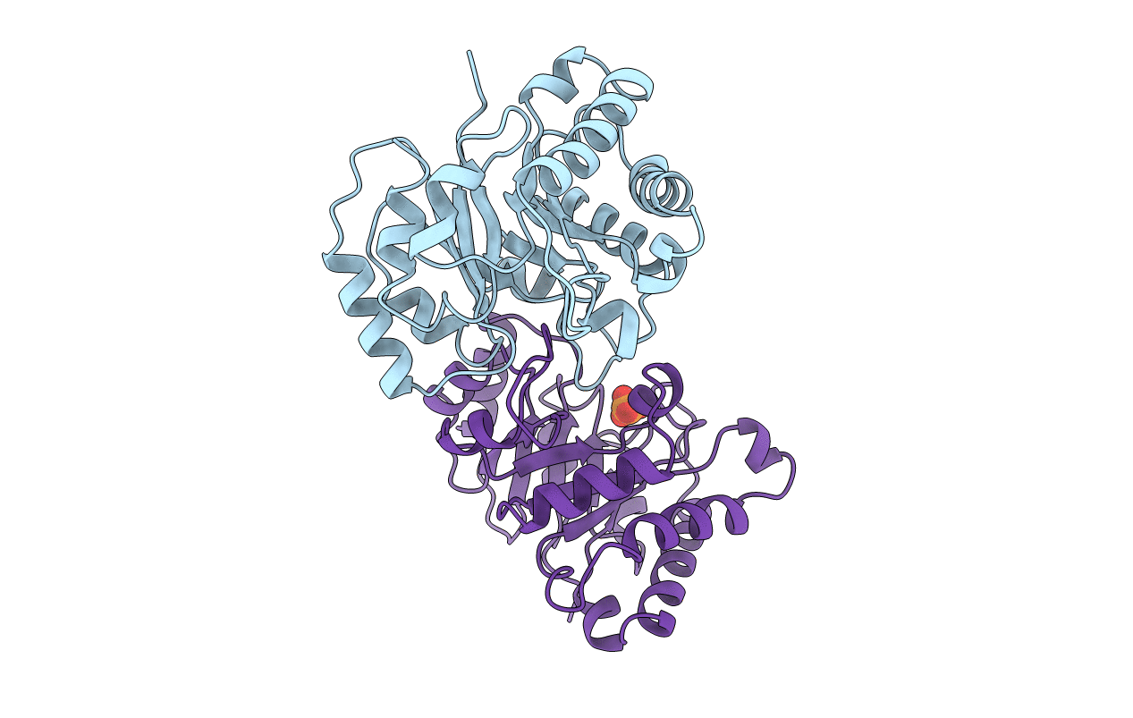
PDB ID:
1AG1
Keywords:
Title:
MONOHYDROGEN PHOSPHATE BINDING TO TRYPANOSOMAL TRIOSEPHOSPHATE ISOMERASE
Biological Source:
Source Organism(s):
Trypanosoma brucei (Taxon ID: 5691)
Method Details:
Experimental Method:
Resolution:
2.36 Å
R-Value Work:
0.15
Space Group:
P 21 21 21