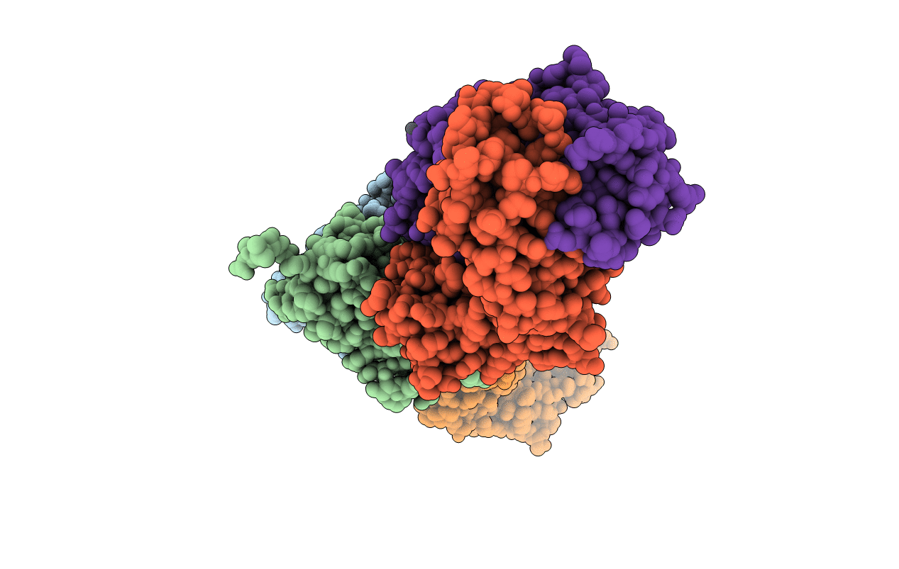
1AFV image
Deposition Date 1997-03-14
Release Date 1997-08-20
Last Version Date 2024-10-09
Entry Detail
PDB ID:
1AFV
Title:
HIV-1 CAPSID PROTEIN (P24) COMPLEX WITH FAB25.3
Biological Source:
Source Organism:
Method Details:
Experimental Method:
Resolution:
3.70 Å
R-Value Free:
0.32
R-Value Work:
0.21
R-Value Observed:
0.21
Space Group:
P 21 2 21
Macromolecular Entities
Polymer Type:polypeptide(L)
Molecule:HUMAN IMMUNODEFICIENCY VIRUS TYPE 1 CAPSID PROTEIN
Gene (Uniprot):gag-pol
Chain IDs:A, B
Chain Length:151
Number of Molecules:2
Biological Source:Human immunodeficiency virus 1
Polymer Type:polypeptide(L)
Molecule:ANTIBODY FAB25.3 FRAGMENT (HEAVY CHAIN)
Gene (Uniprot):Igh
Chain IDs:D (auth: H), F (auth: K)
Chain Length:220
Number of Molecules:2
Biological Source:Mus musculus
Polymer Type:polypeptide(L)
Molecule:ANTIBODY FAB25.3 FRAGMENT (LIGHT CHAIN)
Chain IDs:C (auth: L), E (auth: M)
Chain Length:217
Number of Molecules:2
Biological Source:Mus musculus
Ligand Molecules
Primary Citation

Abstact

X-ray diffraction analysis of a human immunodeficiency virus (HIV-1) capsid (CA) protein shows that each monomer within the dimer consists of seven alpha-helices, five of which are arranged in a coiled coil-like structure. Sequence assignments were made for two of the helices, and tentative connectivity of the remainder of the protein was confirmed by the recent solution structure of a monomeric N-terminal fragment. The C-terminal third of the protein is mostly disordered in the crystal. The longest helices in the coiled coil-like structure are separated by a long, highly antigenic peptide that includes the binding site of an antibody fragment complexed with CA in the crystal. The site of binding of the Fab, the position of the antigenic loop and the site of cleavage between the matrix protein and CA establish the side of the dimer that would be on the exterior of the retroviral core.

Legend

Protein

Chemical

Disease

Primary Citation of related structures