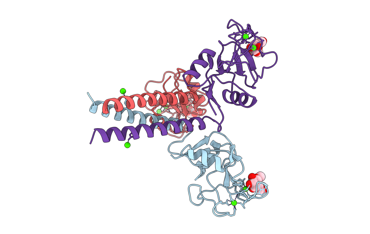
1AFA image
Deposition Date 1995-11-03
Release Date 1996-04-03
Last Version Date 2024-10-30
Entry Detail
PDB ID:
1AFA
Keywords:
Title:
STRUCTURAL BASIS OF GALACTOSE RECOGNITION IN C-TYPE ANIMAL LECTINS
Biological Source:
Source Organism:
Host Organism:
Method Details:
Experimental Method:
Resolution:
2.00 Å
R-Value Free:
0.26
R-Value Work:
0.22
R-Value Observed:
0.22
Space Group:
C 1 2 1
Macromolecular Entities
Structures with similar UniProt ID
Protein Blast
Polymer Type:polypeptide(L)
Molecule:MANNOSE-BINDING PROTEIN-A
Gene (Uniprot):Mbl1
Mutagens:E185Q, N187D, H189W, G190Y, S191G, INS(H192, G193, L194, G195, G196)
Chain IDs:A (auth: 1), B (auth: 2), C (auth: 3)
Chain Length:154
Number of Molecules:3
Biological Source:Rattus norvegicus
Primary Citation
Structural basis of galactose recognition by C-type animal lectins.
J.Biol.Chem. 271 6679 6685 (1996)
PMID: 8636086 DOI: 10.1074/jbc.271.12.6679

Abstact

The asialoglycoprotein receptors and many other C-type (Ca2+-dependent) animal lectins specifically recognize galactose- or N-acetylgalactosamine-terminated oligosaccharides. Analogous binding specificity can be engineered into the homologous rat mannose-binding protein A by changing three amino acids and inserting a glycine-rich loop (Iobst, S. T., and Drickamer, K. (1994) J. Biol. Chem. 269, 15512-15519). Crystal structures of this mutant complexed with beta-methyl galactoside and N-acetylgalactosamine (GalNAc) reveal that as with wild-type mannose-binding proteins, the 3- and 4-OH groups of the sugar directly coordinate Ca2+ and form hydrogen bonds with amino acids that also serve as Ca2+ ligands. The different stereochemistry of the 3- and 4-OH groups in mannose and galactose, combined with a fixed Ca2+ coordination geometry, leads to different pyranose ring locations in the two cases. The glycine-rich loop provides selectivity against mannose by holding a critical tryptophan in a position optimal for packing with the apolar face of galactose but incompatible with mannose binding. The 2-acetamido substituent of GalNAc is in the vicinity of amino acid positions identified by site-directed mutagenesis (Iobst, S. T., and Drickamer, K. (1996) J. Biol. Chem. 271, 6686-6693) as being important for the formation of a GalNAc-selective binding site.

Legend

Protein

Chemical

Disease

Primary Citation of related structures