Deposition Date
1995-12-11
Release Date
1996-06-10
Last Version Date
2024-12-25
Entry Detail
PDB ID:
1AER
Keywords:
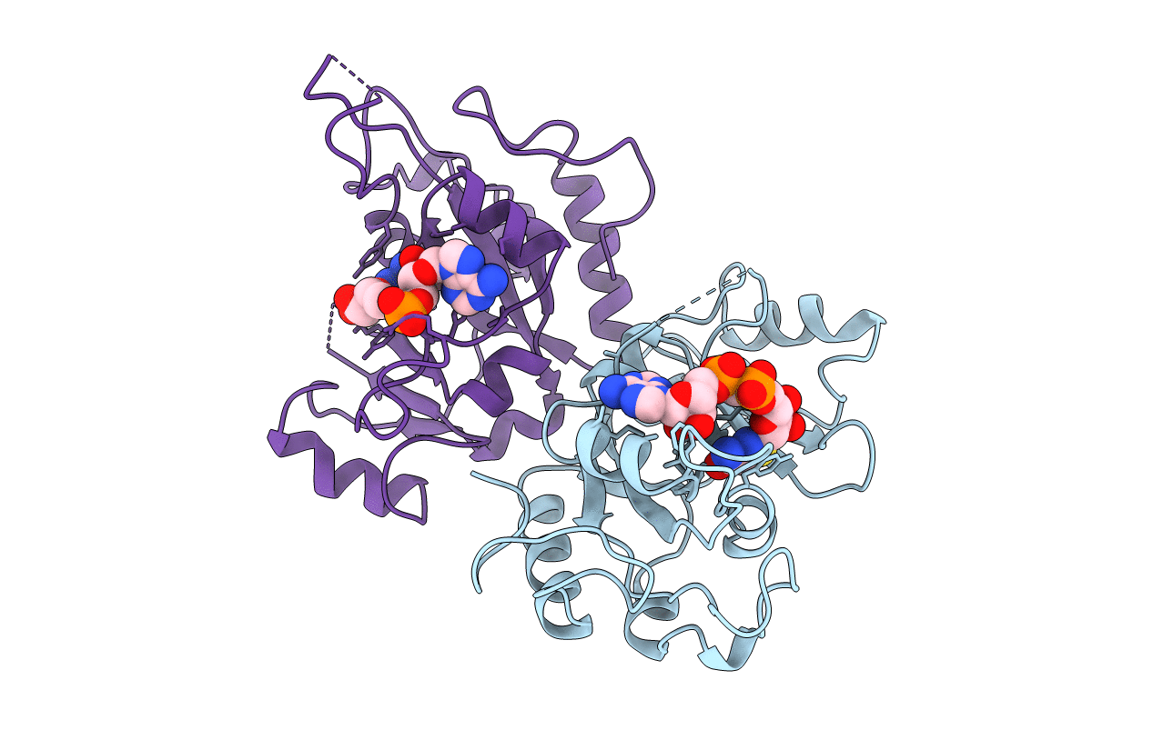
Title:
DOMAIN III OF PSEUDOMONAS AERUGINOSA EXOTOXIN COMPLEXED WITH BETA-TAD
Biological Source:
Source Organism(s):
Pseudomonas aeruginosa (Taxon ID: 287)
Expression System(s):
Method Details:
Experimental Method:
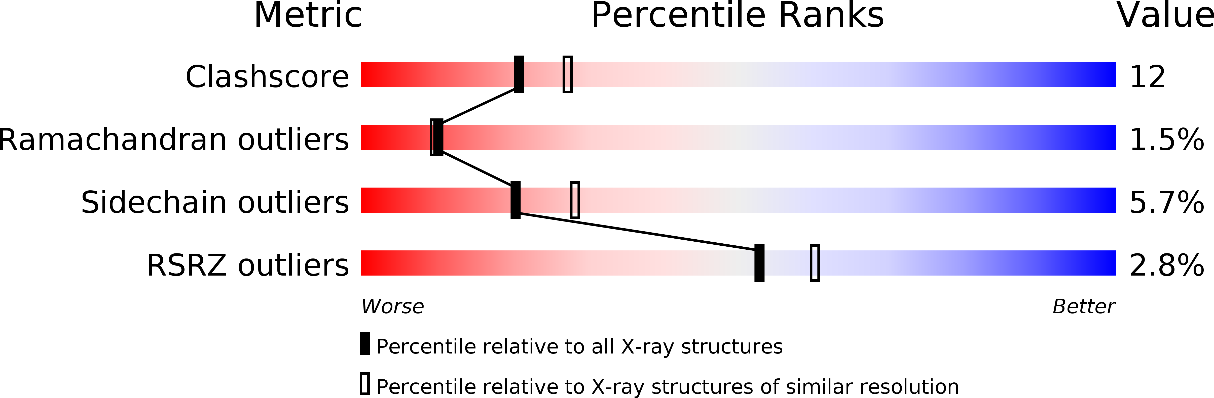
Resolution:
2.30 Å
R-Value Free:
0.28
R-Value Work:
0.19
R-Value Observed:
0.19
Space Group:
P 43 21 2