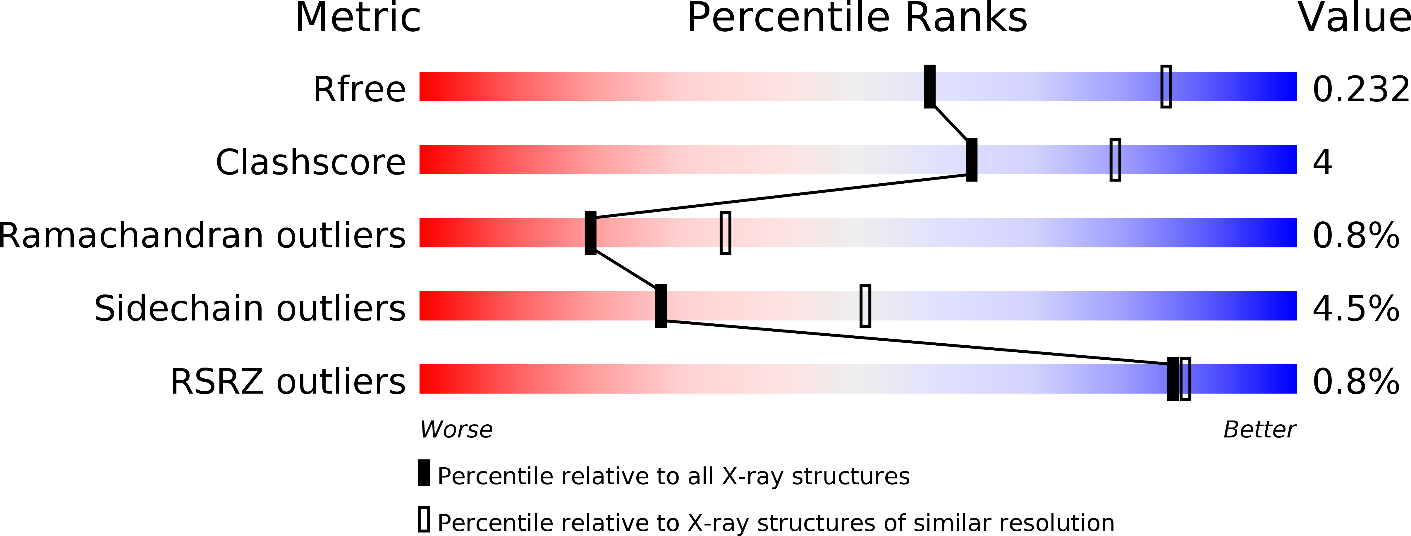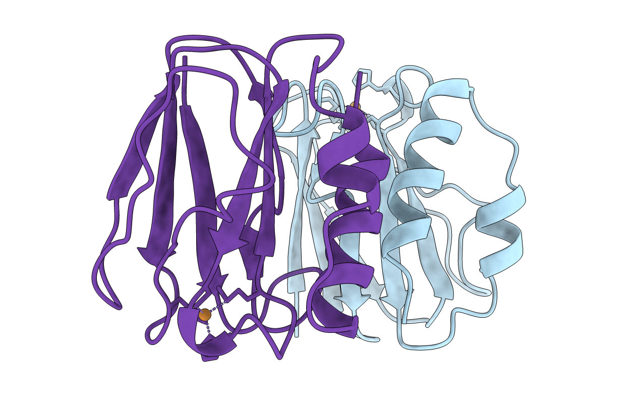
1ADW image
Deposition Date 1997-02-18
Release Date 1997-05-15
Last Version Date 2024-05-22
Entry Detail
PDB ID:
1ADW
Title:
PSEUDOAZURIN
Biological Source:
Source Organism:
Method Details:
Experimental Method:
Resolution:
2.50 Å
R-Value Free:
0.23
R-Value Work:
0.18
R-Value Observed:
0.18
Space Group:
C 1 2 1
Macromolecular Entities
Structures with similar UniProt ID
Protein Blast
Polymer Type:polypeptide(L)
Molecule:PSEUDOAZURIN
Gene (Uniprot):pazS
Chain IDs:A, B
Chain Length:123
Number of Molecules:2
Biological Source:Paracoccus pantotrophus
Ligand Molecules
Primary Citation
Pseudospecific docking surfaces on electron transfer proteins as illustrated by pseudoazurin, cytochrome c550 and cytochrome cd1 nitrite reductase.
Nat.Struct.Biol. 2 975 982 (1995)
PMID: 7583671 DOI: 10.1038/nsb1195-975

Abstact

The structure of pseudoazurin from Thiosphaera pantotropha has been determined and compared to structures of both soluble and membrane-bound periplasmic redox proteins. The results show a matching set of unipolar, but promiscuous, docking motifs based on a positive hydrophobic surface patch on the electron shuttle proteins pseudoazurin and cytochrome c550 and a negative hydrophobic patch on the surface of their known redox partners. The observed electrostatic handedness is argued to be associated with the charge-asymmetry of the membrane-bound components of the redox chain due to von Heijne's 'positives-inside' principle. We propose a 'positives-in-between' rule for electron shuttle proteins, and expect a negative hydrophobic patch to be present on both the highest and lowest redox potential species in a series of electron carriers.

Legend

Protein

Chemical

Disease

Primary Citation of related structures