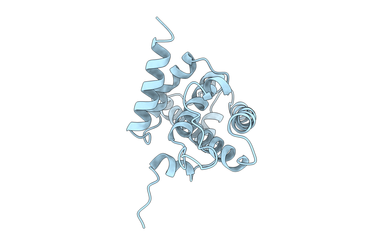
1AD6 image
Deposition Date 1997-02-21
Release Date 1998-08-26
Last Version Date 2024-02-07
Entry Detail
PDB ID:
1AD6
Title:
DOMAIN A OF HUMAN RETINOBLASTOMA TUMOR SUPPRESSOR
Biological Source:
Source Organism:
Homo sapiens (Taxon ID: 9606)
Method Details:
Experimental Method:
Resolution:
2.30 Å
R-Value Free:
0.24
R-Value Work:
0.20
Space Group:
P 21 21 21
Macromolecular Entities
Structures with similar UniProt ID
Protein Blast
Polymer Type:polypeptide(L)
Molecule:RETINOBLASTOMA TUMOR SUPPRESSOR
Gene (Uniprot):RB1
Chain IDs:A
Chain Length:185
Number of Molecules:1
Biological Source:Homo sapiens
Primary Citation
Structural similarity between the pocket region of retinoblastoma tumour suppressor and the cyclin-box.
Nat.Struct.Biol. 4 390 395 (1997)
PMID: 9145110 DOI: 10.1038/nsb0597-390

Abstact

The pocket region of retinoblastoma tumour suppressor (Rb) is essential for tumour suppressing activity. The Rb pocket is primarily composed of two domains, A and B. We have determined the X-ray crystal structure of domain A (residues 378-562) at 2.3 A resolution. Domain A consists of nine alpha-helices. The overall arrangement of helices in domain A is remarkably similar to the cyclin-box folds found in the crystal structures of cyclin A and TFIIB. This structure, along with domain B which is predicted to be homologous to the cyclin-box, suggests that the Rb pocket is composed of two cyclin-box fold domains. We present the structural/functional features of the Rb pocket, and the potential binding region for cellular or viral proteins within domain A.

Legend

Protein

Chemical

Disease

Primary Citation of related structures