1AD4 image
Entry Detail
PDB ID:
1AD4
Keywords:
Title:
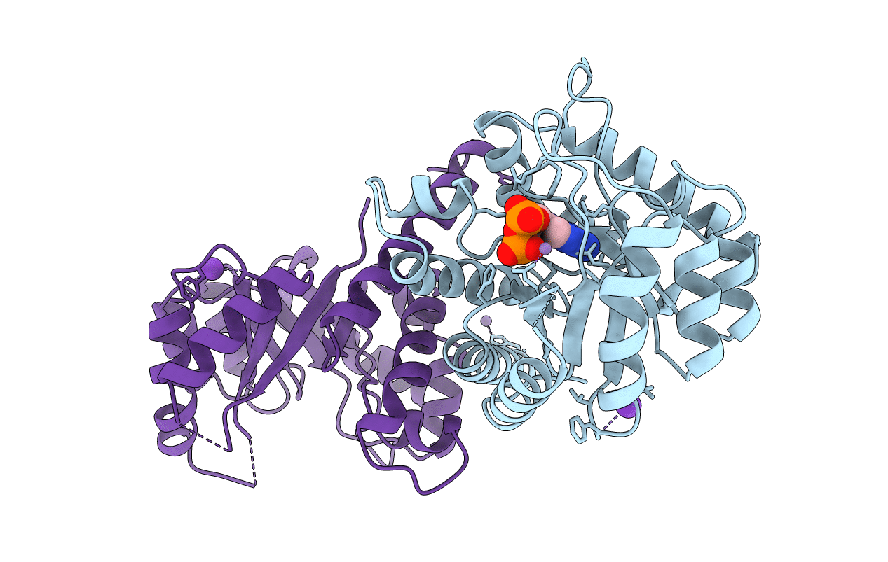
DIHYDROPTEROATE SYNTHETASE COMPLEXED WITH OH-CH2-PTERIN-PYROPHOSPHATE FROM STAPHYLOCOCCUS AUREUS
Biological Source:
Source Organism:
PDB Version:
Deposition Date:
1997-02-20
Release Date:
1998-04-29
Method Details:
Experimental Method:
Resolution:
2.40 Å
R-Value Work:
0.17
R-Value Observed:
0.17
Space Group:
P 1 21 1
Macromolecular Entities
Polymer Type:polypeptide(L)
Description:DIHYDROPTEROATE SYNTHETASE
Chain IDs:A, B
Chain Length:266
Number of Molecules:2
Biological Source:Staphylococcus aureus
Primary Citation
Structure and function of the dihydropteroate synthase from Staphylococcus aureus.
J.Mol.Biol. 268 21 30 (1997)
PMID: 9149138 DOI: 10.1006/jmbi.1997.0944

Abstact

The gene encoding the dihydropteroate synthase of staphylococcus aureus has been cloned, sequenced and expressed in Escherichia coli. The protein has been purified for biochemical characterization and X-ray crystallographic studies. The enzyme is a dimer in solution, has a steady state kinetic mechanism that suggests random binding of the two substrates and half-site reactivity. The crystal structure of apo-enzyme and a binary complex with the substrate analogue hydroxymethylpterin pyrophosphate were determined at 2.2 A and 2.4 A resolution, respectively. The enzyme belongs to the group of "TIM-barrel" proteins and crystallizes as a non-crystallographic dimer. Only one molecule of the substrate analogue bound per dimer in the crystal. Sequencing of nine sulfonamide-resistant clinical isolates has shown that as many as 14 residues could be involved in resistance development. The residues are distributed over the surface of the protein, which defies a simple interpretation of their roles in resistance. Nevertheless, the three-dimensional structure of the substrate analogue binary complex could give important insight into the molecular mechanism of this enzyme.

Legend

Protein

Chemical

Disease

Primary Citation of related structures