
Deposition Date
1993-07-19
Release Date
1994-01-31
Last Version Date
2024-05-22
Entry Detail
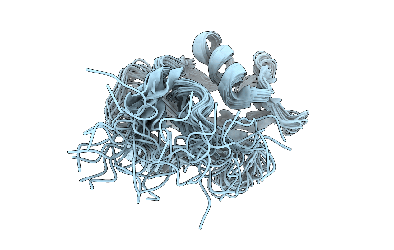
PDB ID:
1AB2
Keywords:
Title:
THREE-DIMENSIONAL SOLUTION STRUCTURE OF THE SRC HOMOLOGY 2 DOMAIN OF C-ABL
Biological Source:
Source Organism(s):
Homo sapiens (Taxon ID: 9606)
Method Details:
Experimental Method:
Conformers Submitted:
20
