Deposition Date
1998-04-06
Release Date
1998-07-15
Last Version Date
2024-04-10
Entry Detail
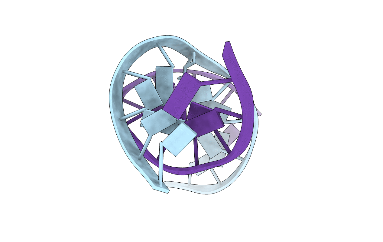
PDB ID:
1A9I
Keywords:
Title:
APYRIMIDINIC DNA WITH BOUND WATER AT THE DAMAGED SITE, ALPHA FORM, NMR, 1 STRUCTURE
Method Details:
Experimental Method:
Conformers Calculated:
100
Conformers Submitted:
1
Selection Criteria:
AVERAGE OF 5 STRUCTURES THAT THE NOESY BACKCALCULATION AGREES WITH EXPERIMENTAL NOESY