
Deposition Date
1998-04-15
Release Date
1998-10-21
Last Version Date
2024-10-09
Entry Detail
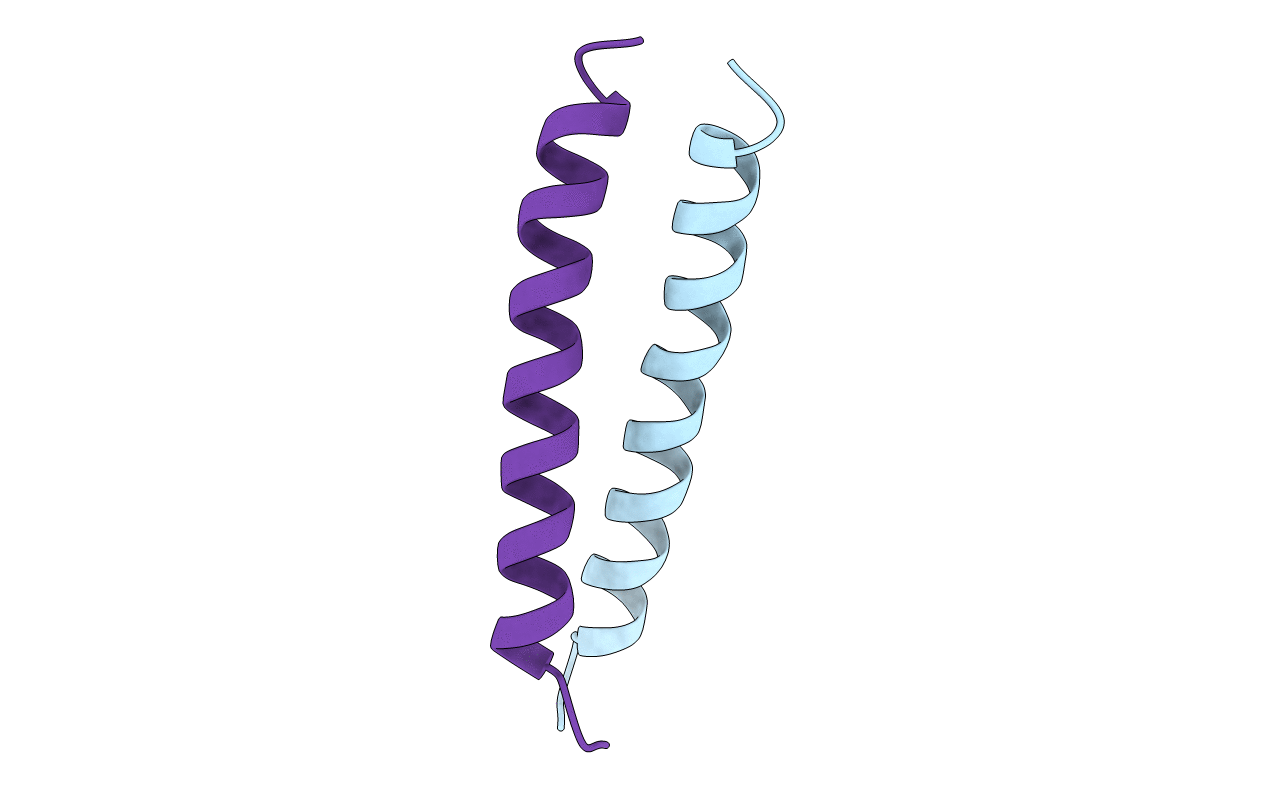
PDB ID:
1A93
Keywords:
Title:
NMR SOLUTION STRUCTURE OF THE C-MYC-MAX HETERODIMERIC LEUCINE ZIPPER, NMR, MINIMIZED AVERAGE STRUCTURE
Biological Source:
Source Organism(s):
Homo sapiens (Taxon ID: 9606)
Mus musculus (Taxon ID: 10090)
Mus musculus (Taxon ID: 10090)
Method Details:
Experimental Method:
Conformers Calculated:
40
Conformers Submitted:
1
Selection Criteria:
LEAST RESTRAINT VIOLATION ONLY
