Deposition Date
1998-03-24
Release Date
1998-07-15
Last Version Date
2024-05-22
Entry Detail
PDB ID:
1A8G
Keywords:
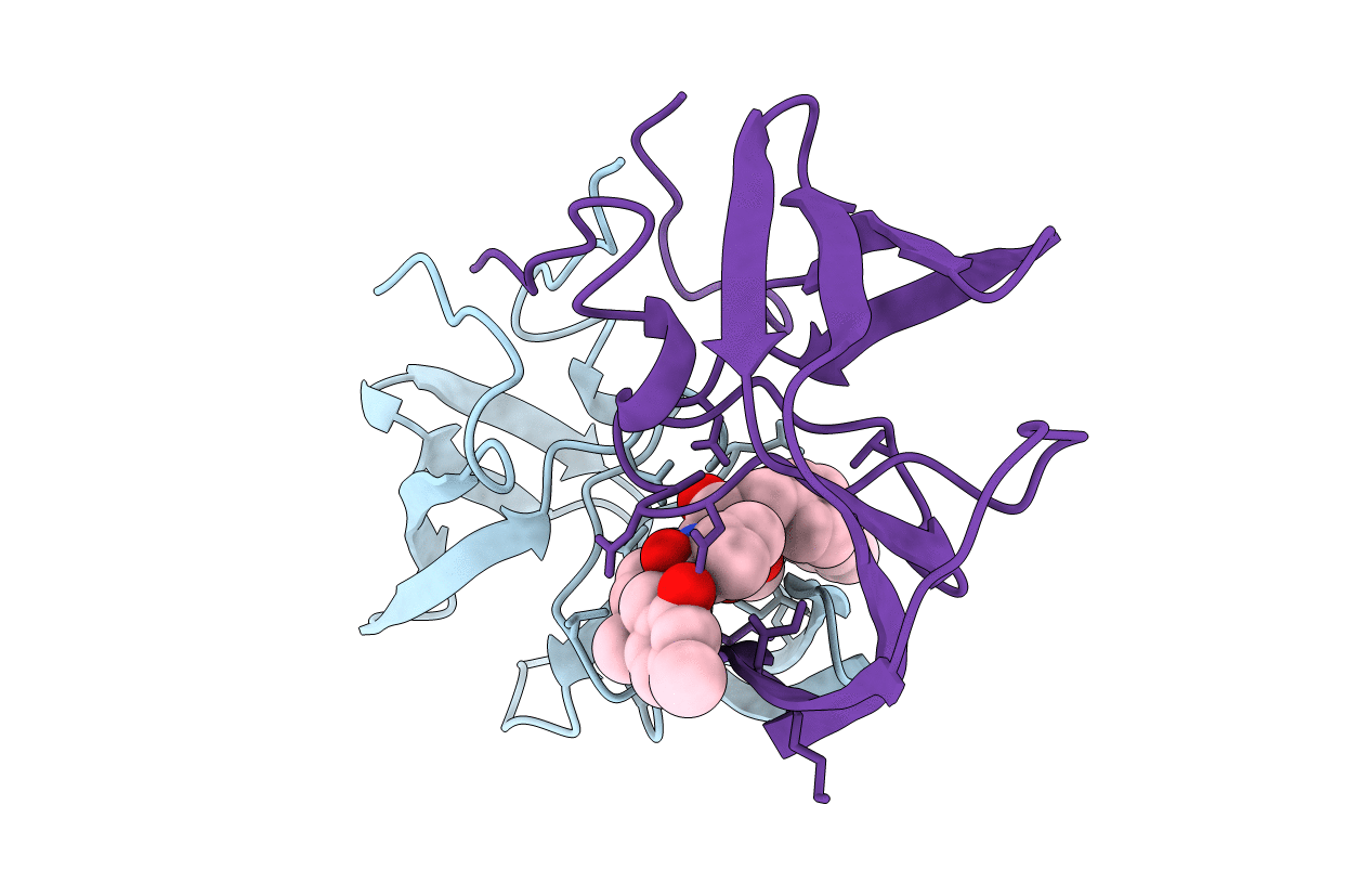
Title:
HIV-1 PROTEASE IN COMPLEX WITH SDZ283-910
Biological Source:
Source Organism:
Human immunodeficiency virus 1 (Taxon ID: 11676)
Host Organism:
Method Details:
Experimental Method:
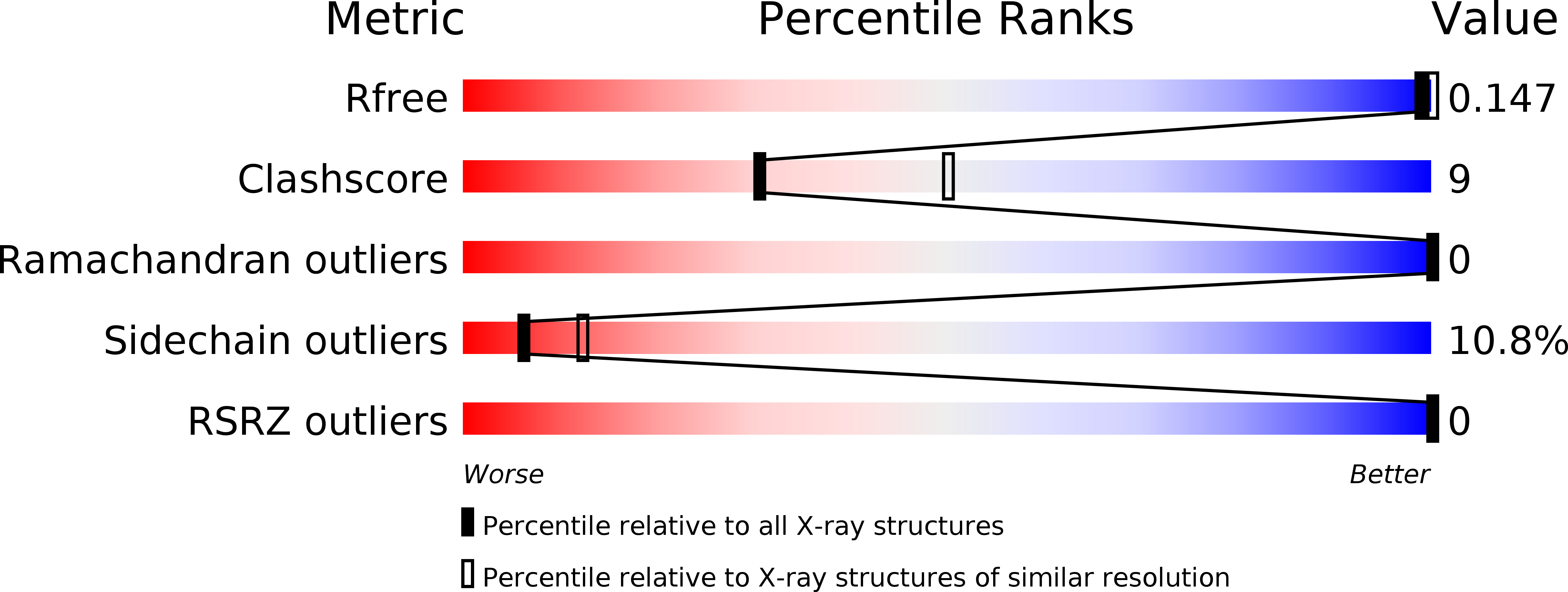
Resolution:
2.50 Å
R-Value Free:
0.24
R-Value Work:
0.15
R-Value Observed:
0.15
Space Group:
P 61