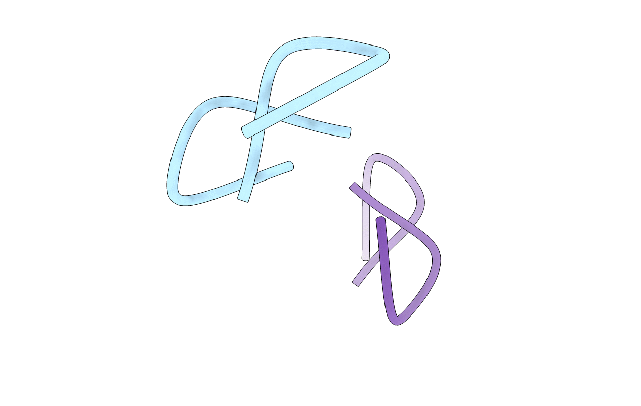
1A7Z image
Deposition Date 1998-03-19
Release Date 1999-03-23
Last Version Date 2024-07-10
Entry Detail
PDB ID:
1A7Z
Keywords:
Title:
CRYSTAL STRUCTURE OF ACTINOMYCIN Z3
Biological Source:
Source Organism:
Method Details:
Experimental Method:
Resolution:
0.95 Å
R-Value Free:
0.09
R-Value Observed:
0.08
Space Group:
P 21 21 21
Macromolecular Entities
Polymer Type:polypeptide(L)
Molecule:ACTINOMYCIN Z3
Chain IDs:A, B
Chain Length:11
Number of Molecules:2
Biological Source:STREPTOMYCES FRADIAE
Ligand Molecules
Peptide-like Molecules
PRD_000009
Primary Citation
Crystal Structures of Actinomycin D and Actinomycin Z3.
Angew.Chem.Int.Ed.Engl. 37 2381 2384 (1998)
PMID: 29710967 DOI: 10.1002/(SICI)1521-3773(19980918)37:17<2381::AID-ANIE2381>3.0.CO;2-L

Abstact

Untwinned single crystals of the actinomycins D and Z3 that diffracted to atomic resolution could be obtained for the first time. Low-temperature data collection and a new ab initio method for solving the structures led to precise crystal structures which showed, for example, that the unit cell of actinomycin D contains three molecules, two of which are present in the form of a hydrogen-bridged dimer related by a pseudo-twofold axis (see picture).

Legend

Protein

Chemical

Disease

Primary Citation of related structures