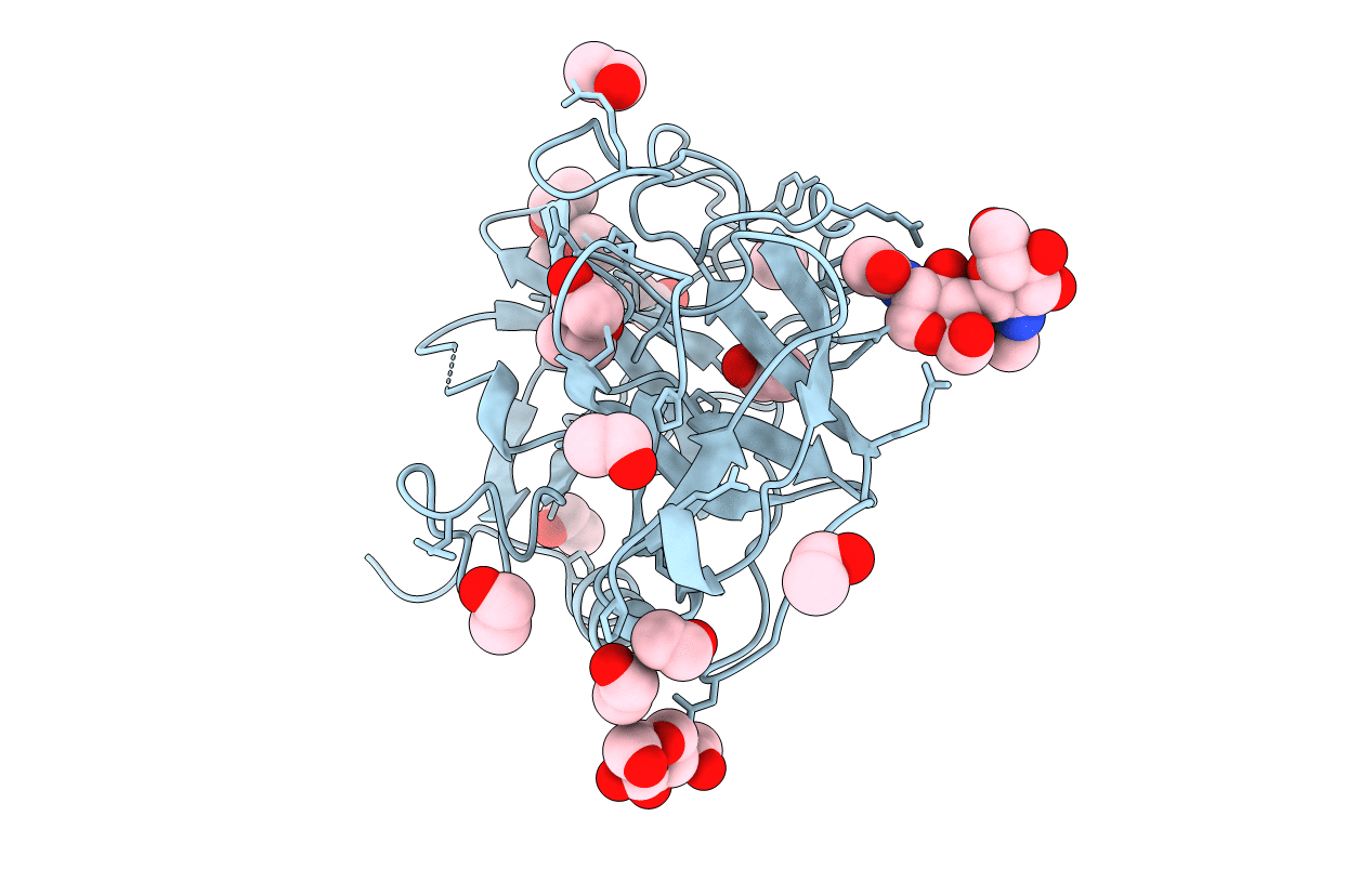
1A7S image
Deposition Date 1998-03-17
Release Date 1999-03-23
Last Version Date 2024-11-06
Entry Detail
PDB ID:
1A7S
Title:
ATOMIC RESOLUTION STRUCTURE OF HBP
Biological Source:
Source Organism:
Homo sapiens (Taxon ID: 9606)
Host Organism:
Method Details:
Experimental Method:
Resolution:
1.12 Å
R-Value Free:
0.18
R-Value Observed:
0.15
Space Group:
P 21 21 21
Macromolecular Entities
Polymer Type:polypeptide(L)
Molecule:HEPARIN BINDING PROTEIN
Gene (Uniprot):AZU1
Chain IDs:A
Chain Length:225
Number of Molecules:1
Biological Source:Homo sapiens
Modified Residue
Compound ID Chain ID Parent Comp ID Details 2D Image
ASN A ASN GLYCOSYLATION SITE
Primary Citation

Abstact

Crystals of human heparin binding protein (HBP) diffract to 1.1 A when flash-frozen at 120 K. The atomic resolution structure has been refined anisotropically using SHELXL96. The final model of HBP consists of 221 amino-acid residues of 225 possible, three glycosylation units, one chloride ion, 15 precipitant ethanol molecules and 323 water molecules. The structure is refined to a final crystallographic R factor of 15.9% and Rfree(5%) of 18.9% using all data. A putative protein kinase C activation site has been identified, involving residues 113-120. The structure is compared to the previously determined 2.3 A resolution structure of HBP.

Legend

Protein

Chemical

Disease

Primary Citation of related structures