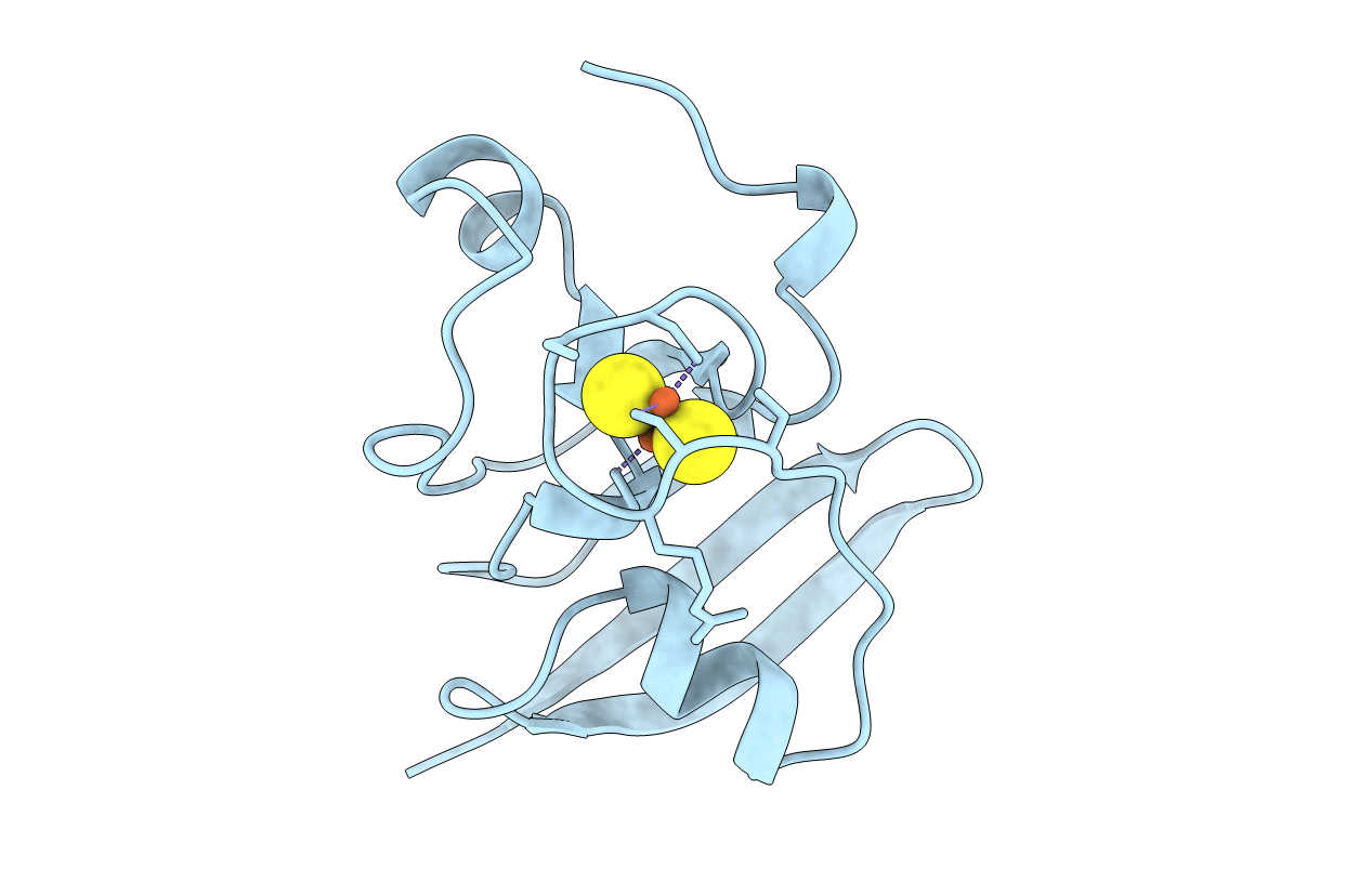
1A70 image
Entry Detail
PDB ID:
1A70
Title:
SPINACH FERREDOXIN
Biological Source:
Source Organism:
Host Organism:
PDB Version:
Deposition Date:
1998-03-19
Release Date:
1998-11-25
Method Details:
Experimental Method:
Resolution:
1.70 Å
R-Value Free:
0.20
R-Value Work:
0.18
R-Value Observed:
0.19
Space Group:
P 21 21 21
Macromolecular Entities
Polymer Type:polypeptide(L)
Description:FERREDOXIN
Mutations:E92K
Chain IDs:A
Chain Length:97
Number of Molecules:1
Biological Source:Spinacia oleracea
Ligand Molecules
Primary Citation
Structure of the mutant E92K of [2Fe-2S] ferredoxin I from Spinacia oleracea at 1.7 A resolution.
Acta Crystallogr.,Sect.D 54 1353 1358 (1998)
PMID: 10089511 DOI: 10.1107/S0907444998005137

Abstact

Ferredoxin I (Fd I) from Spinacia oleracea is composed of 97 amino-acid residues and a [2Fe-2S] cluster. The crystal structure of the E92K mutant of Fd I was solved by molecular replacement and refined to an R factor of 19.6% for 11755 reflections at 1.7 A resolution. The overall structure and the active centre of spinach Fd is highly conserved with respect to ferredoxins of known structure. The E92K mutation appears to disturb a hydrogen-bond network which stabilizes the loop bearing the [2Fe-2S] cluster. This observation provides a rationale for the reduced electron-transfer efficiency displayed by the E92K mutant. Inspection of the crystal packing reveals that the side chain of Lys92 is engaged in an intermolecular interaction with Asp26 of a symmetry-related molecule. This feature may explain why only the mutant E92K and not wild-type Fd I could be successfully crystallized.

Legend

Protein

Chemical

Disease

Primary Citation of related structures