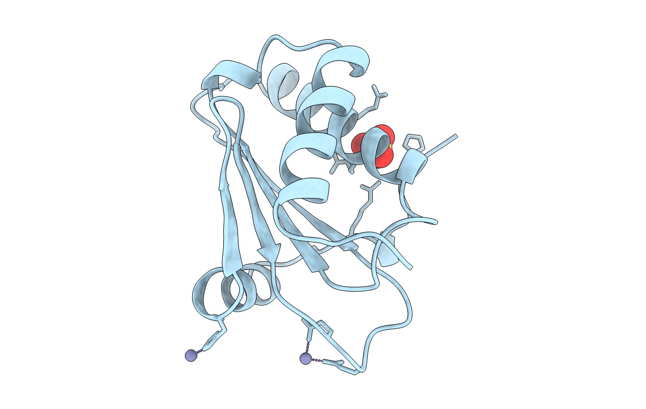
1A6F image
Entry Detail
PDB ID:
1A6F
Keywords:
Title:
RNASE P PROTEIN FROM BACILLUS SUBTILIS
Biological Source:
Source Organism:
Host Organism:
PDB Version:
Deposition Date:
1998-02-24
Release Date:
1999-03-23
Method Details:
Experimental Method:
Resolution:
2.60 Å
R-Value Free:
0.31
R-Value Work:
0.20
R-Value Observed:
0.20
Space Group:
P 64
Macromolecular Entities
Polymer Type:polypeptide(L)
Description:RIBONUCLEASE P PROTEIN
Mutations:ALA 2, HIS 3, LEU 4 ARE INSERTED BETWEEN MET 1 AND LYS 2
Chain IDs:A
Chain Length:119
Number of Molecules:1
Biological Source:Bacillus subtilis
Primary Citation
Ribonuclease P protein structure: evolutionary origins in the translational apparatus.
Science 280 752 755 (1998)
PMID: 9563955 DOI: 10.1126/science.280.5364.752

Abstact

The crystal structure of Bacillus subtilis ribonuclease P protein is reported at 2.6 angstroms resolution. This protein binds to ribonuclease P RNA to form a ribonucleoprotein holoenzyme with optimal catalytic activity. Mutagenesis and biochemical data indicate that an unusual left-handed betaalphabeta crossover connection and a large central cleft in the protein form conserved RNA binding sites; a metal binding loop may comprise a third RNA binding site. The unusual topology is partly shared with ribosomal protein S5 and the ribosomal translocase elongation factor G, which suggests evolution from a common RNA binding ancestor in the primordial translational apparatus.

Legend

Protein

Chemical

Disease

Primary Citation of related structures