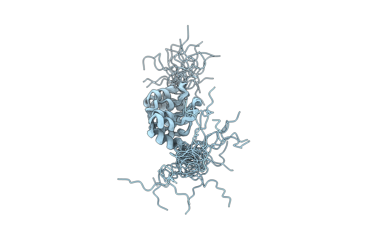
1A5E image
Deposition Date 1998-02-13
Release Date 1999-08-13
Last Version Date 2024-05-22
Entry Detail
PDB ID:
1A5E
Keywords:
Title:
SOLUTION NMR STRUCTURE OF TUMOR SUPPRESSOR P16INK4A, 18 STRUCTURES
Biological Source:
Source Organism:
Homo sapiens (Taxon ID: 9606)
Method Details:
Experimental Method:
Conformers Calculated:
90
Conformers Submitted:
18
Selection Criteria:
CLOSEST TO MEAN STRUCTURE WHICH SHOWS GOOD AGREEMENT WITH THE CONSTRAINTS. NONE OF THE CONSTRAINTS SHOW NOE VIOLATION BIGGER THAN 0.5 A AND DIHEDRAL ANGLE VIOLATION BIGGER THAN 5 DEGREE.
Macromolecular Entities
Structures with similar UniProt ID
Protein Blast
Polymer Type:polypeptide(L)
Molecule:TUMOR SUPPRESSOR P16INK4A
Gene (Uniprot):CDKN2A
Chain IDs:A
Chain Length:156
Number of Molecules:1
Biological Source:Homo sapiens
Ligand Molecules
Primary Citation
Tumor suppressor p16INK4A: determination of solution structure and analyses of its interaction with cyclin-dependent kinase 4.
Mol.Cell 1 421 431 (1998)
PMID: 9660926 DOI: 10.1016/S1097-2765(00)80042-8

Abstact

The solution structure of the tumor suppressor p16INK4A has been determined by NMR, and important recognition regions of both cdk4 and p16INK4A have been identified. The tertiary structure of p16INK4A contains four helix-turn-helix motifs linked by three loops. Twelve tumorigenic mutants of p16INK4A have been constructed and analyzed for their structure and activity, and new mutants have been designed rationally. A fragment of 58 residues at the N terminus of cdk4 important for p16INK4A binding has been identified. The importance of this region was further verified by mutational analysis of cdk4. These results and docking experiments have been used to assess possible modes of binding between p16INK4A and cdk4.

Legend

Protein

Chemical

Disease

Primary Citation of related structures