1A4X image
Deposition Date 1998-02-08
Release Date 1998-08-05
Last Version Date 2024-05-22
Entry Detail
PDB ID:
1A4X
Title:
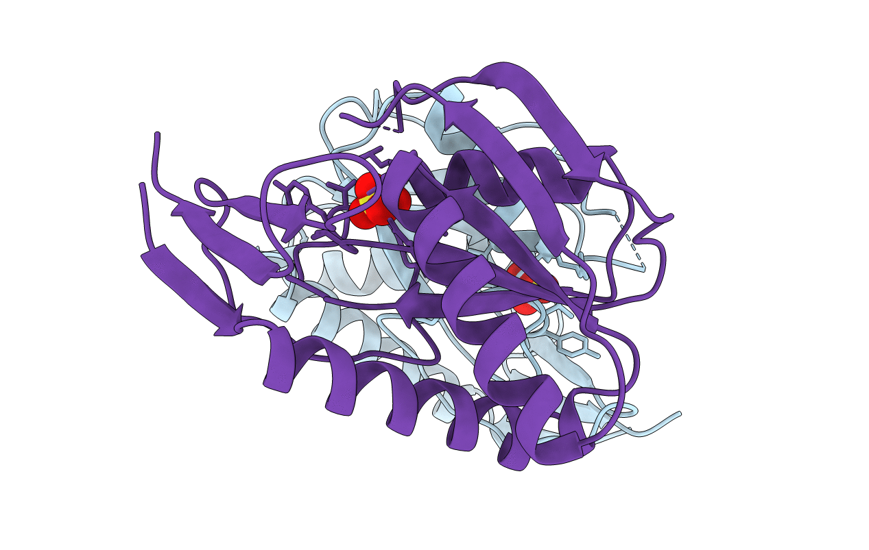
PYRR, THE BACILLUS SUBTILIS PYRIMIDINE BIOSYNTHETIC OPERON REPRESSOR, HEXAMERIC FORM
Biological Source:
Source Organism:
Host Organism:
Method Details:
Experimental Method:
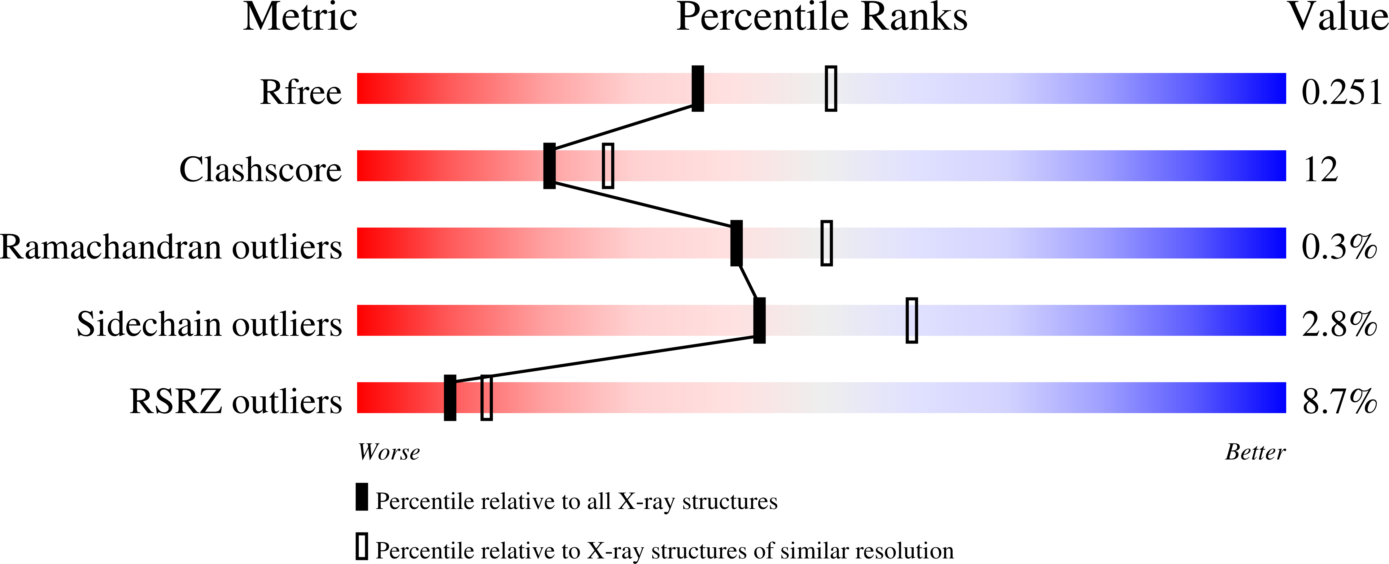
Resolution:
2.30 Å
R-Value Free:
0.24
R-Value Work:
0.21
R-Value Observed:
0.21
Space Group:
H 3 2
Macromolecular Entities
Polymer Type:polypeptide(L)
Molecule:PYRIMIDINE OPERON REGULATORY PROTEIN PYRR
Gene (Uniprot):pyrR
Chain IDs:A, B
Chain Length:181
Number of Molecules:2
Biological Source:Bacillus subtilis
Ligand Molecules
Primary Citation
Adaptation of an enzyme to regulatory function: structure of Bacillus subtilis PyrR, a pyr RNA-binding attenuation protein and uracil phosphoribosyltransferase.
Structure 6 337 350 (1998)
PMID: 9551555 DOI: 10.1016/S0969-2126(98)00036-7

Abstact

BACKGROUND The expression of pyrimidine nucleotide biosynthetic (pyr) genes in Bacillus subtilis is regulated by transcriptional attenuation. The PyrR attenuation protein binds to specific sites in pyr mRNA, allowing the formation of downstream terminator structures. UMP and 5-phosphoribosyl-1-pyrophosphate (PRPP), a nucleotide metabolite, are co-regulators with PyrR. The smallest RNA shown to bind tightly to PyrR is a 28-30 nucleotide stem-loop that contains a purine-rich bulge and a putative-GNRA tetraloop. PyrR is also a uracil phosphoribosyltransferase (UPRTase), although the relationship between enzymatic activity and RNA recognition is unclear, and the UPRTase activity of PyrR is not physiologically significant in B. subtilis. Elucidating the role of PyrR structural motifs in UMP-dependent RNA binding is an important step towards understanding the mechanism of pyr transcriptional attenuation. RESULTS The 1.6 A crystal structure of B. subtilis PyrR has been determined by multiwavelength anomalous diffraction, using a Sm co-crystal. As expected, the structure of PyrR is homologous to those proteins of the large type I PRTase structural family; it is most similar to hypoxanthine-guanine-xanthine PRTase (HGXPRTase). The PyrR dimer differs from other PRTase dimers, suggesting it may have evolved specifically for RNA binding. A large, basic, surface at the dimer interface is an obvious RNA-binding site and uracil specificity is probably provided by hydrogen bonds from mainchain and sidechain atoms in the hood subdomain. These models of RNA and UMP binding are consistent with biological data. CONCLUSIONS The B. subtilis protein PyrR has adapted the substrate- and product-binding capacities of a PRTase, probably an HGXPRTase, producing a new regulatory function in which the substrate and product are co-regulators of transcription termination. The structure is consistent with the idea that PyrR regulatory function is independent of catalytic activity, which is likely to be extremely low under physiological conditions.

Legend

Protein

Chemical

Disease

Primary Citation of related structures