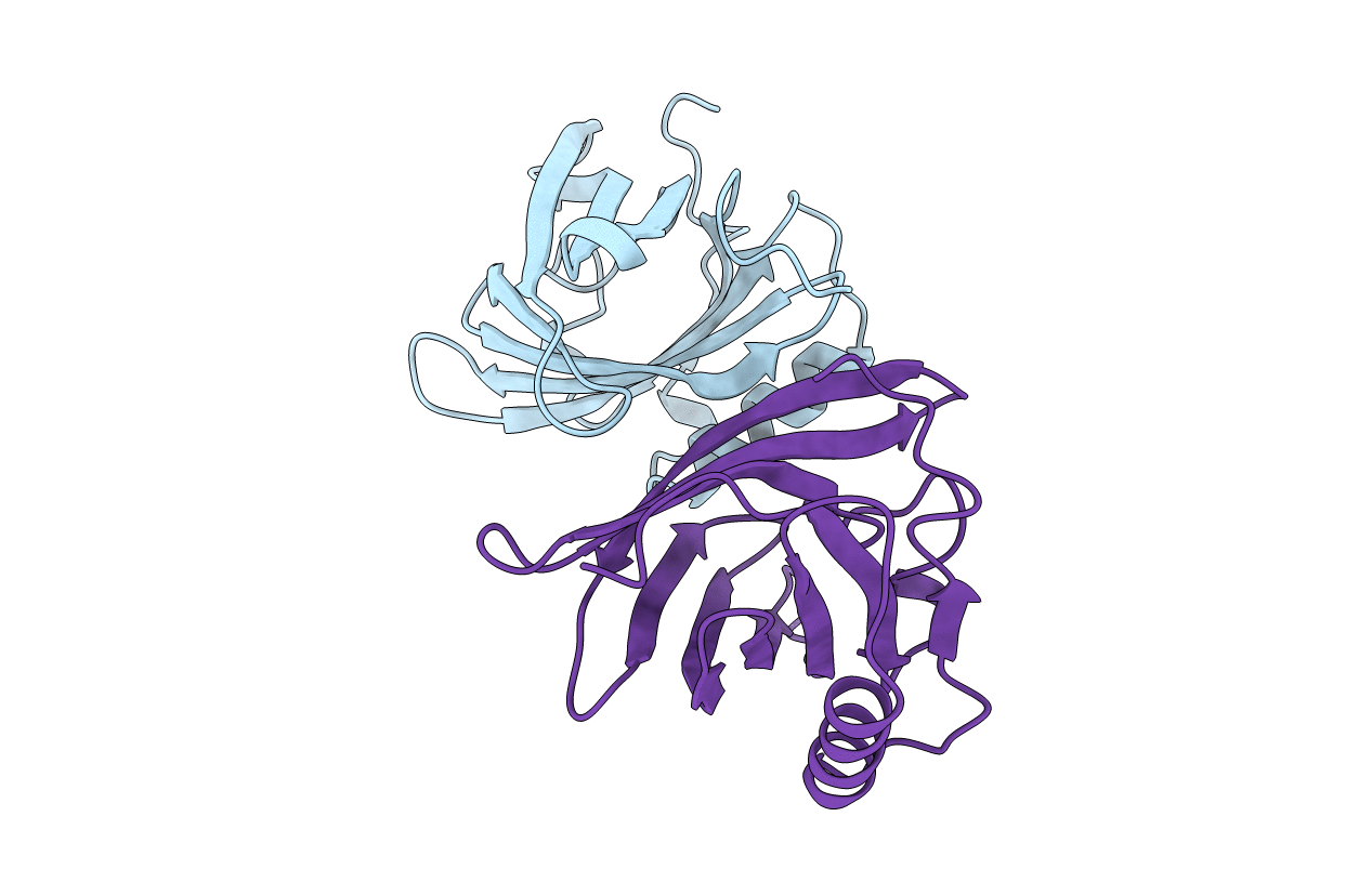
1A3Y image
Deposition Date 1998-01-27
Release Date 1999-02-16
Last Version Date 2024-10-30
Entry Detail
PDB ID:
1A3Y
Keywords:
Title:
ODORANT BINDING PROTEIN FROM NASAL MUCOSA OF PIG
Biological Source:
Source Organism:
Sus scrofa (Taxon ID: 9823)
Method Details:
Experimental Method:
Resolution:
2.25 Å
R-Value Free:
0.24
R-Value Work:
0.18
R-Value Observed:
0.18
Space Group:
P 21 21 21
Macromolecular Entities
Polymer Type:polypeptide(L)
Molecule:ODORANT BINDING PROTEIN
Chain IDs:A, B
Chain Length:149
Number of Molecules:2
Biological Source:Sus scrofa
Primary Citation
The structure of the monomeric porcine odorant binding protein sheds light on the domain swapping mechanism.
Biochemistry 37 7913 7918 (1998)
PMID: 9609684 DOI: 10.1021/bi980179e

Abstact

The X-ray structure of the porcine odorant binding protein (OBPp) was determined at 2.25 A resolution. This lipocalin is a monomer and is devoid of naturally occurring bound ligand, contrary to what was observed in the case of bovine OBP [Tegoni, M., et al. (1996) Nat. Struct. Biol. 3, 863-867; Bianchet, M. A., et al. (1996) Nat. Struct. Biol. 3, 934-939]. In this latter protein, a dimer without any disulfide bridges, domain swapping was found to occur between the beta- and alpha-domains. A single Gly (121) insertion was found in OBPp when it was compared to OBPb, which may prevent domain swapping from taking place. The presence of a disulfide bridge between the OBPp beta- and alpha-domains (cysteines 63 and 155) may lock the resulting fold in a nonswapped monomeric conformation. Comparisons with other OBPs indicate that the two cysteines involved in the OBPp disulfide bridge are conserved in the sequence, suggesting that OBPp may be considered a prototypic OBP fold, and not OBPb.

Legend

Protein

Chemical

Disease

Primary Citation of related structures