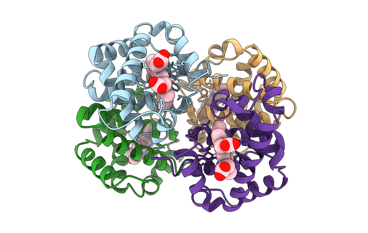
1A3O image
Deposition Date 1998-01-22
Release Date 1998-04-29
Last Version Date 2024-02-07
Entry Detail
PDB ID:
1A3O
Title:
ARTIFICIAL MUTANT (ALPHA Y42H) OF DEOXY HEMOGLOBIN
Biological Source:
Source Organism:
Homo sapiens (Taxon ID: 9606)
Method Details:
Experimental Method:
Resolution:
1.80 Å
R-Value Free:
0.22
R-Value Work:
0.17
Space Group:
P 1 21 1
Macromolecular Entities
Structures with similar UniProt ID
Protein Blast
Polymer Type:polypeptide(L)
Molecule:HEMOGLOBIN (ALPHA CHAIN)
Gene (Uniprot):HBA1, HBA2
Mutagens:CHAIN A, C, Y42H
Chain IDs:A, C
Chain Length:141
Number of Molecules:2
Biological Source:Homo sapiens
Structures with similar UniProt ID
Protein Blast
Polymer Type:polypeptide(L)
Molecule:HEMOGLOBIN (BETA CHAIN)
Gene (Uniprot):HBB
Mutagens:CHAIN A, C, Y42H
Chain IDs:B, D
Chain Length:146
Number of Molecules:2
Biological Source:Homo sapiens
Ligand Molecules
Primary Citation
The structures of deoxy human haemoglobin and the mutant Hb Tyralpha42His at 120 K.
Acta Crystallogr.,Sect.D 56 805 811 (2000)
PMID: 10930827 DOI: 10.1107/S0907444900006387

Abstact

The structures of deoxy human haemoglobin and an artificial mutant (Tyralpha42-->His) have been solved at 120 K. While overall agreement between these structures and others in the PDB is very good, certain side chains are found to be shifted, absent from the electron-density map or in different rotamers. Non-crystallographic symmetry (NCS) is very well obeyed in the native protein, but not around the site of the changed residue in the mutant. NCS is also not obeyed by the water molecule invariably found in the alpha-chain haem pocket in room-temperature crystal structures of haemoglobin. At 120 K, this water molecule disappears from one alpha chain in the asymmetric unit but not the other.

Legend

Protein

Chemical

Disease

Primary Citation of related structures