Deposition Date
1998-01-21
Release Date
1999-03-16
Last Version Date
2024-02-07
Entry Detail
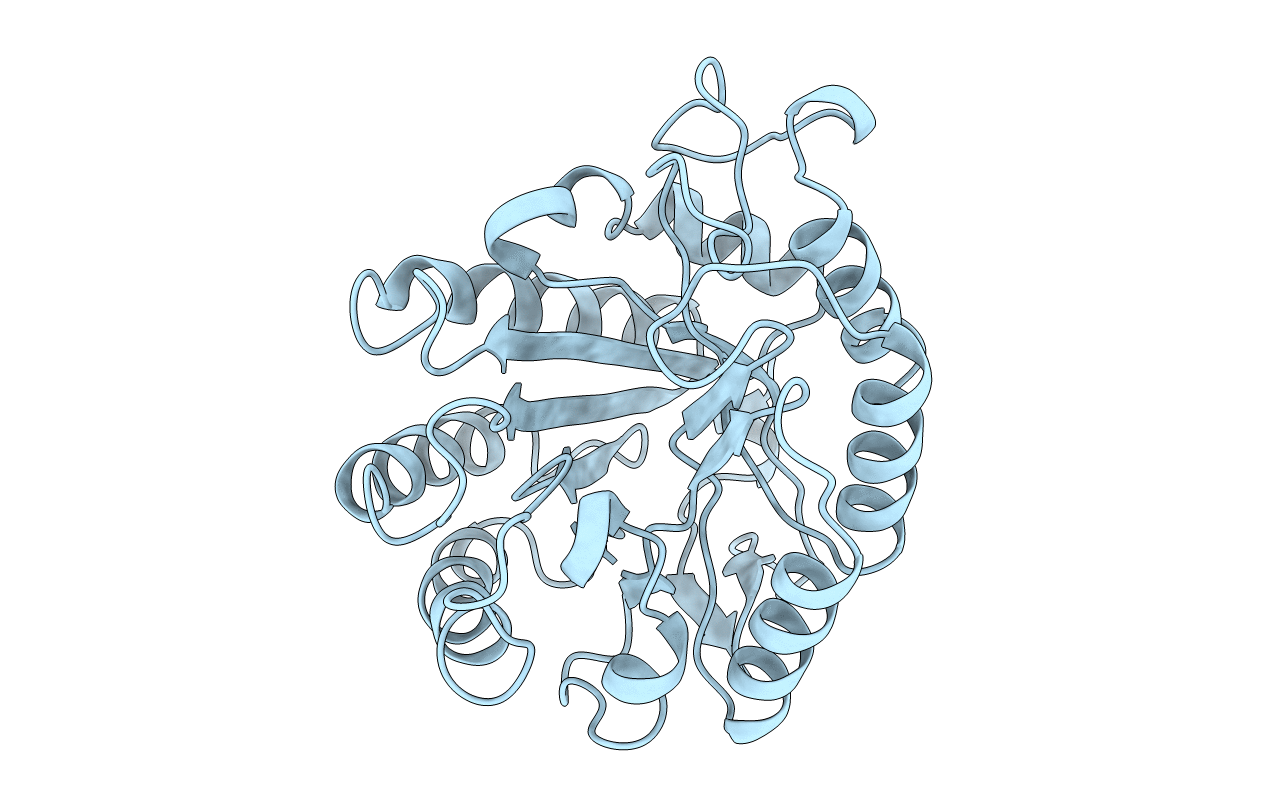
PDB ID:
1A3H
Keywords:
Title:
ENDOGLUCANASE CEL5A FROM BACILLUS AGARADHERANS AT 1.6A RESOLUTION
Biological Source:
Source Organism(s):
Bacillus agaradhaerens (Taxon ID: 76935)
Expression System(s):
Method Details:
Experimental Method:
Resolution:
1.57 Å
R-Value Free:
0.17
R-Value Work:
0.14
Space Group:
P 21 21 21