Deposition Date
1998-01-21
Release Date
1998-04-29
Last Version Date
2024-11-06
Entry Detail
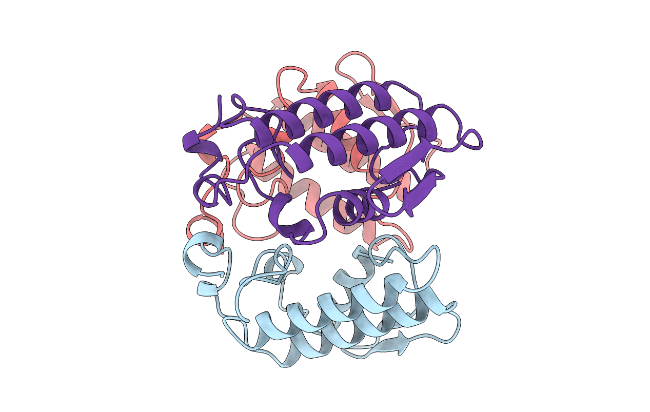
PDB ID:
1A3F
Keywords:
Title:
PHOSPHOLIPASE A2 (PLA2) FROM NAJA NAJA VENOM
Biological Source:
Method Details:
Experimental Method:
Resolution:
2.65 Å
R-Value Free:
0.26
R-Value Work:
0.21
R-Value Observed:
0.21
Space Group:
P 21 21 21