1A0R image
Deposition Date 1997-12-05
Release Date 1998-12-30
Last Version Date 2024-10-16
Entry Detail
PDB ID:
1A0R
Title:
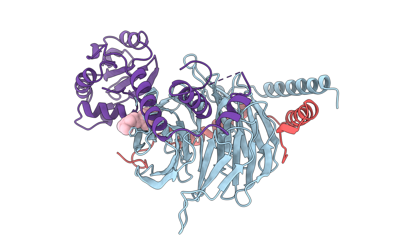
HETEROTRIMERIC COMPLEX OF PHOSDUCIN/TRANSDUCIN BETA-GAMMA
Biological Source:
Source Organism:
Bos taurus (Taxon ID: 9913)
Method Details:
Experimental Method:
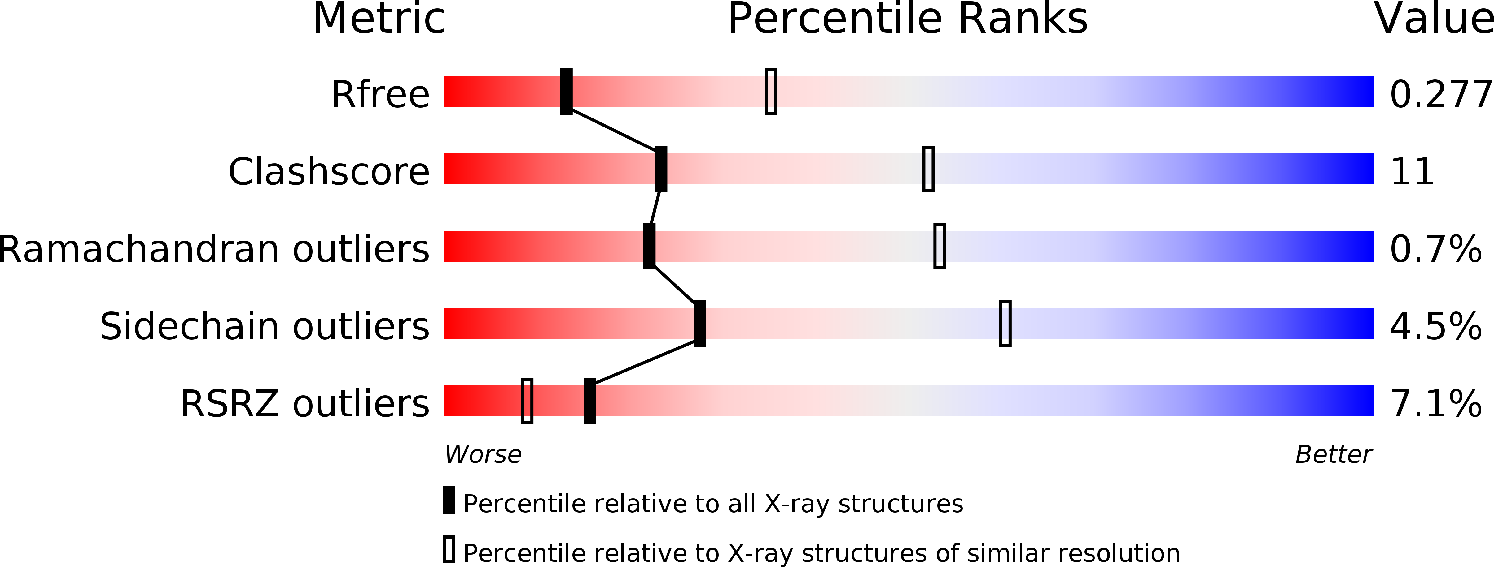
Resolution:
2.80 Å
R-Value Free:
0.26
R-Value Work:
0.22
R-Value Observed:
0.22
Space Group:
P 21 21 21
Macromolecular Entities
Structures with similar UniProt ID
Protein Blast
Polymer Type:polypeptide(L)
Molecule:TRANSDUCIN (BETA SUBUNIT)
Chain IDs:A (auth: B)
Chain Length:340
Number of Molecules:1
Biological Source:Bos taurus
Structures with similar UniProt ID
Protein Blast
Polymer Type:polypeptide(L)
Molecule:TRANSDUCIN (GAMMA SUBUNIT)
Gene (Uniprot):GNGT1
Chain IDs:B (auth: G)
Chain Length:65
Number of Molecules:1
Biological Source:Bos taurus
Structures with similar UniProt ID
Protein Blast
Polymer Type:polypeptide(L)
Molecule:PHOSDUCIN
Gene (Uniprot):PDC
Chain IDs:C (auth: P)
Chain Length:245
Number of Molecules:1
Biological Source:Bos taurus
Ligand Molecules
Primary Citation
Phosducin induces a structural change in transducin beta gamma.
Structure 6 1007 1019 (1998)
PMID: 9739091 DOI: 10.1016/S0969-2126(98)00102-6

Abstact

BACKGROUND Phosducin binds tightly to the beta gamma subunits (Gt beta gamma) of the heterotrimeric G protein transducin, preventing Gt beta gamma reassociation with Gt alpha-GDP and thereby inhibiting the G-protein cycle. Phosducin-like proteins appear to be widely distributed and may play important roles in regulating many heterotrimeric G-protein signaling pathways. RESULTS The 2.8 A crystal structure of a complex of bovine retinal phosducin with Gt beta gamma shows how the two domains of phosducin cover one side and the top of the seven-bladed beta propeller of Gt beta gamma. The binding of phosducin induces a distinct structural change in the beta propeller of Gt beta gamma, such that a small cavity opens up between blades 6 and 7. Electron density in this cavity has been assigned to the farnesyl moiety of the gamma subunit. CONCLUSIONS beta gamma subunits of heterotrimeric G proteins can exist in two distinct conformations. In the R (relaxed) state, corresponding to the structure of the free beta gamma or the structure of beta gamma in the alpha beta gamma heterotrimer, the hydrophobic farnesyl moiety of the gamma subunit is exposed, thereby mediating membrane association. In the T (tense) state, as observed in the phosducin-Gt beta gamma structure, the farnesyl moiety of the gamma subunit is effectively buried in the cavity formed between blades 6 and 7 of the beta subunit. Binding of phosducin to Gt beta gamma induces the formation of this cavity, resulting in a switch from the R to the T conformation. This sequesters beta gamma from the membrane to the cytosol and turns off the signal-transduction cascade. Regulation of this membrane association/dissociation switch of Gt beta gamma by phosducin may be a general mechanism for attenuation of G protein coupled signal transduction cascades.

Legend

Protein

Chemical

Disease

Primary Citation of related structures