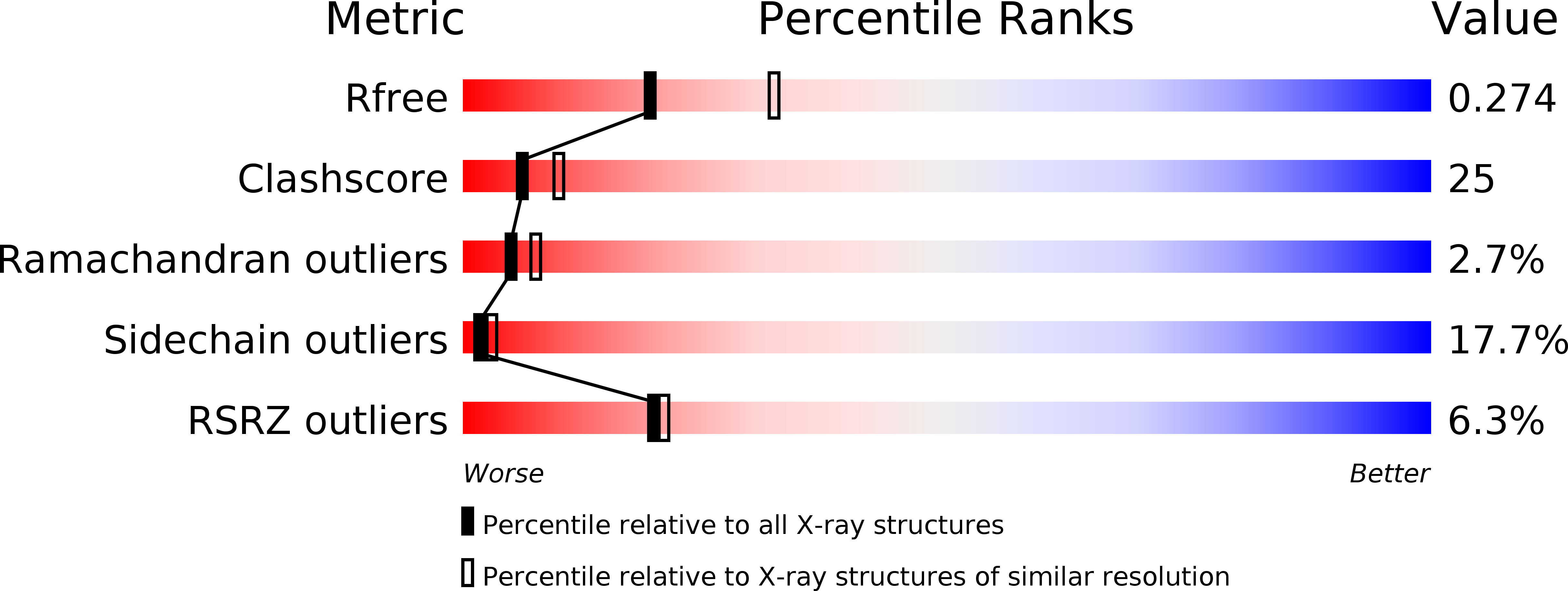
1A0P image
Deposition Date 1997-12-05
Release Date 1998-03-18
Last Version Date 2024-02-07
Entry Detail
PDB ID:
1A0P
Title:
SITE-SPECIFIC RECOMBINASE, XERD
Biological Source:
Source Organism:
Escherichia coli (Taxon ID: 562)
Host Organism:
Method Details:
Experimental Method:
Resolution:
2.50 Å
R-Value Free:
0.28
R-Value Work:
0.22
Space Group:
P 65
Macromolecular Entities
Structures with similar UniProt ID
Protein Blast
Polymer Type:polypeptide(L)
Molecule:SITE-SPECIFIC RECOMBINASE XERD
Gene (Uniprot):xerD
Chain IDs:A
Chain Length:290
Number of Molecules:1
Biological Source:Escherichia coli
Primary Citation
Crystal structure of the site-specific recombinase, XerD.
EMBO J. 16 5178 5187 (1997)
PMID: 9311978 DOI: 10.1093/emboj/16.17.5178

Abstact

The structure of the site-specific recombinase, XerD, that functions in circular chromosome separation, has been solved at 2.5 A resolution and reveals that the protein comprises two domains. The C-terminal domain contains two conserved sequence motifs that are located in similar positions in the structures of XerD, lambda and HP1 integrases. However, the extreme C-terminal regions of the three proteins, containing the active site tyrosine, are very different. In XerD, the arrangement of active site residues supports a cis cleavage mechanism. Biochemical evidence for DNA bending is encompassed in a model that accommodates extensive biochemical and genetic data, and in which the DNA is wrapped around an alpha-helix in a manner similar to that observed for CAP complexed with DNA.

Legend

Protein

Chemical

Disease

Primary Citation of related structures