Deposition Date
1997-12-03
Release Date
1999-01-13
Last Version Date
2024-10-30
Entry Detail
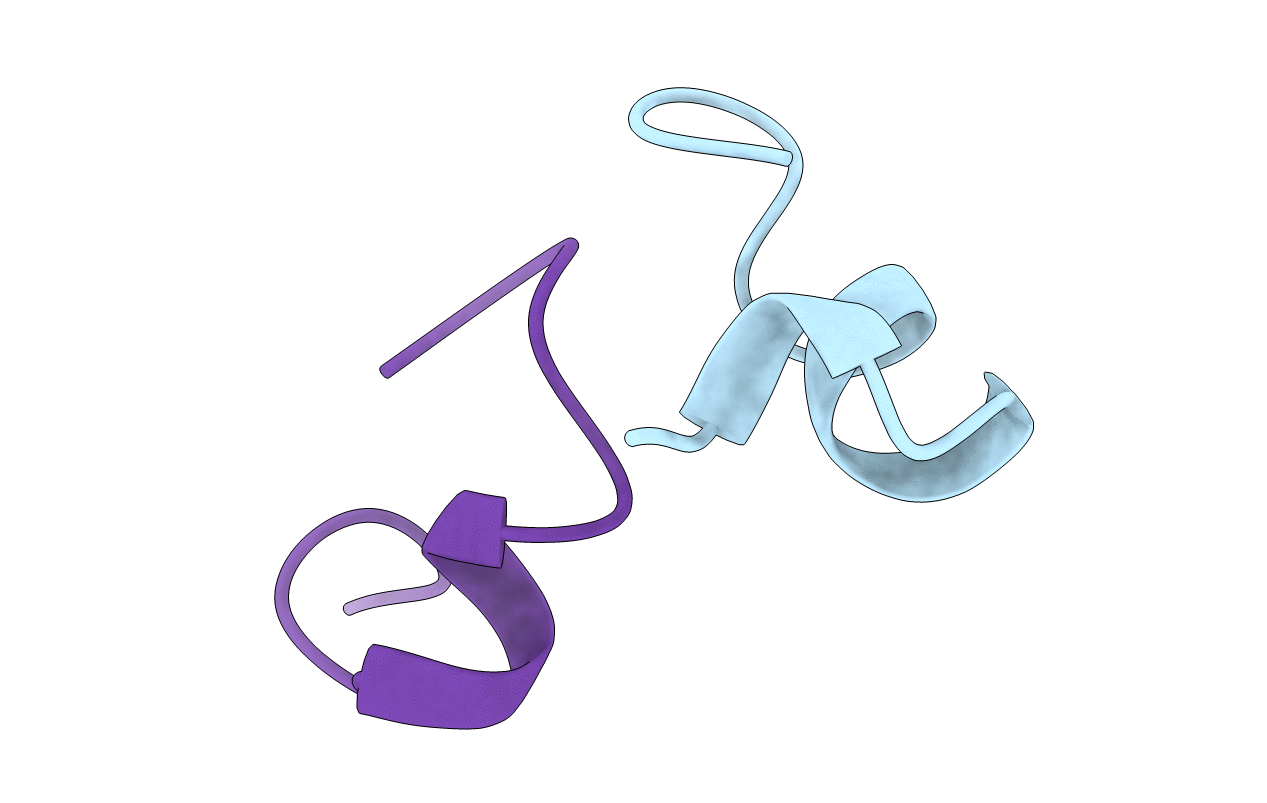
PDB ID:
1A0M
Keywords:
Title:
1.1 ANGSTROM CRYSTAL STRUCTURE OF A-CONOTOXIN [TYR15]-EPI
Biological Source:
Source Organism(s):
Conus episcopatus (Taxon ID: 88764)
Method Details:
Experimental Method:
Resolution:
1.10 Å
R-Value Free:
0.17
R-Value Work:
0.16
R-Value Observed:
0.16
Space Group:
I 4