1A05 image
Deposition Date 1997-12-09
Release Date 1998-06-17
Last Version Date 2024-05-22
Entry Detail
PDB ID:
1A05
Keywords:
Title:
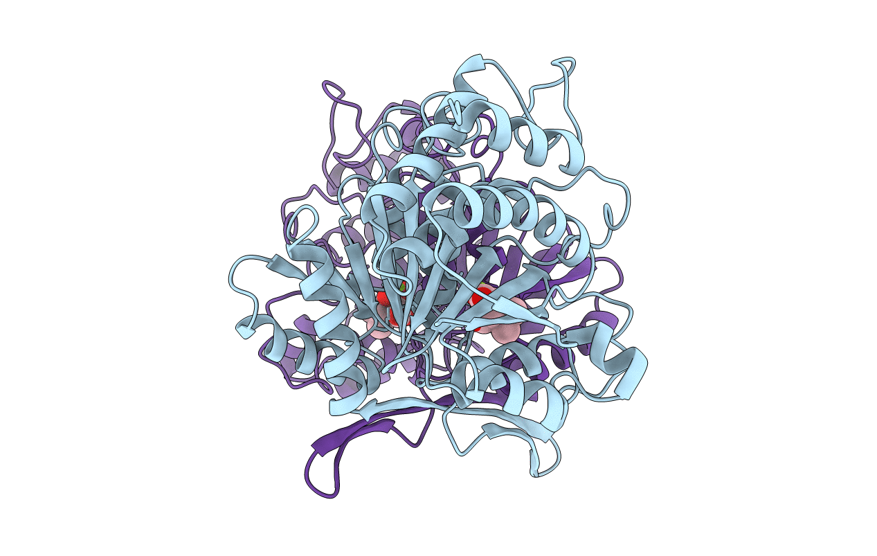
CRYSTAL STRUCTURE OF THE COMPLEX OF 3-ISOPROPYLMALATE DEHYDROGENASE FROM THIOBACILLUS FERROOXIDANS WITH 3-ISOPROPYLMALATE
Biological Source:
Source Organism:
Host Organism:
Method Details:
Experimental Method:
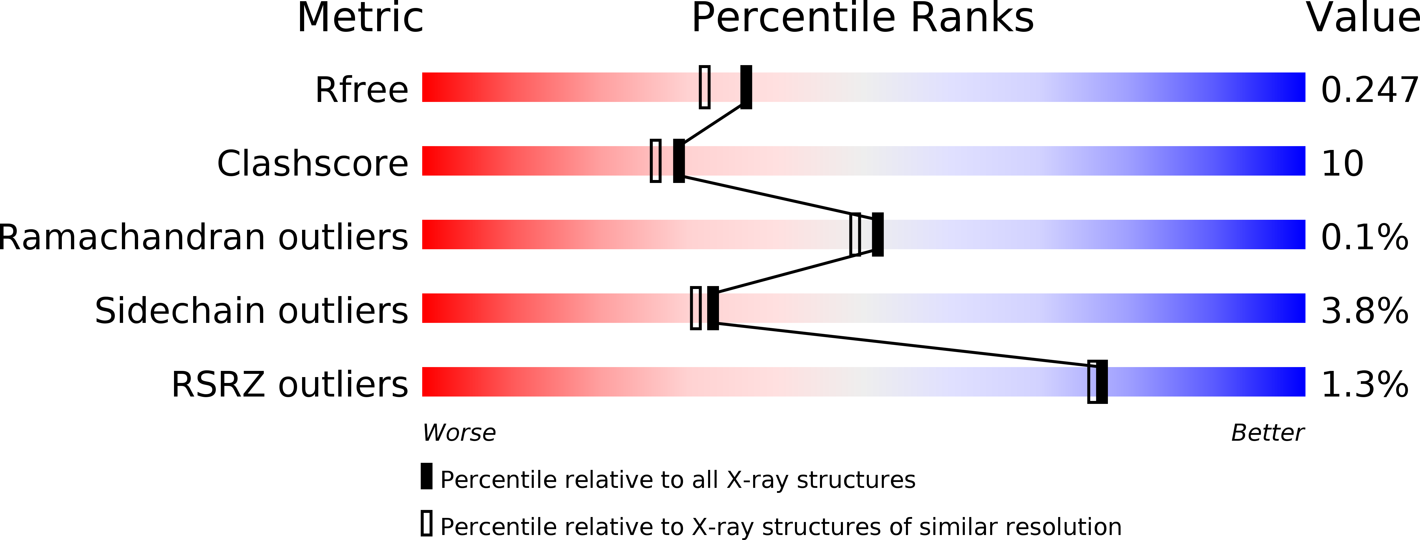
Resolution:
2.00 Å
R-Value Free:
0.27
R-Value Work:
0.19
R-Value Observed:
0.19
Space Group:
P 21 21 2
Macromolecular Entities
Structures with similar UniProt ID
Protein Blast
Polymer Type:polypeptide(L)
Molecule:3-ISOPROPYLMALATE DEHYDROGENASE
Gene (Uniprot):leuB
Chain IDs:A, B
Chain Length:358
Number of Molecules:2
Biological Source:Acidithiobacillus ferrooxidans
Primary Citation
Structure of 3-isopropylmalate dehydrogenase in complex with 3-isopropylmalate at 2.0 A resolution: the role of Glu88 in the unique substrate-recognition mechanism.
Structure 6 971 982 (1998)
PMID: 9739088 DOI: 10.1016/S0969-2126(98)00099-9

Abstact

BACKGROUND 3-Isopropylmalate dehydrogenase (IPMDH) and isocitrate dehydrogenase (ICDH) belong to a unique family of bifunctional decarboxylating dehydrogenases. Although the ICDH dimer catalyzes its reaction under a closed conformation, known structures of the IPMDH dimer (without substrate) adopt a fully open or a partially closed form. Considering the similarity in the catalytic mechanism, the IPMDH dimer must be in a fully closed conformation during the reaction. A large conformational change should therefore occur upon substrate binding. RESULTS We have determined the crystal structure of IPMDH from Thiobacillus ferrooxidans (Tf) complexed with 3-isopropylmalate (IPM) at 2.0 A resolution by the molecular replacement method. The structure shows a fully closed conformation and the substrate-binding site is quite similar to that of ICDH except for a region around the gamma-isopropyl group. The gamma group is recognized by a unique hydrophobic pocket, which includes Glu88, Leu91 and Leu92 from subunit 1 and Val193' from subunit 2. CONCLUSIONS A large movement of domain 1 is induced by substrate binding, which results in the formation of the hydrophobic pocket for the gamma-isopropyl moiety of IPM. A glutamic acid in domain 1, Glu88, participates in the formation of the hydrophobic pocket. The C beta and C gamma atoms of Glu88 interact with the gamma-isopropyl moiety of IPM and are central to the recognition of substrate. The acidic tip of Glu88 is likely to interact with the nicotinamide mononucleotide (NMN) ribose of NAD+ in the ternary complex. This structure clearly explains the substrate specificity of IPMDH.

Legend

Protein

Chemical

Disease

Primary Citation of related structures