186D image
Deposition Date 1994-08-22
Release Date 1994-11-01
Last Version Date 2024-05-22
Entry Detail
PDB ID:
186D
Keywords:
Title:
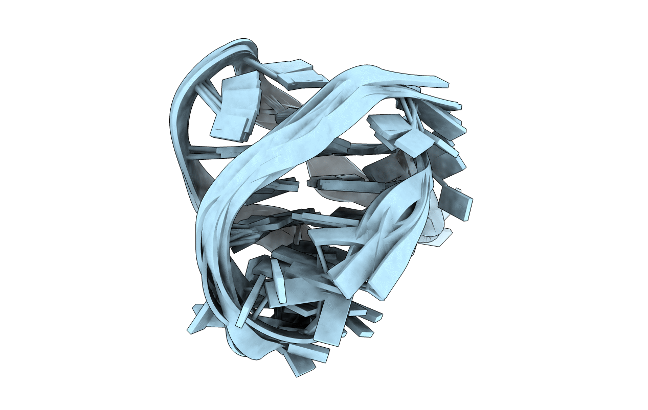
SOLUTION STRUCTURE OF THE TETRAHYMENA TELOMERIC REPEAT D(T2G4)4 G-TETRAPLEX
Biological Source:
Source Organism:
Method Details:
Experimental Method:
Conformers Submitted:
7
Macromolecular Entities
Polymer Type:polydeoxyribonucleotide
Molecule:DNA (5'-D(*TP*TP*GP*GP*GP*GP*TP*TP*GP*GP*GP*GP*TP*TP*GP*GP*GP*GP*TP*TP*GP*GP*GP*G)-3')
Chain IDs:A
Chain Length:24
Number of Molecules:1
Biological Source:
Ligand Molecules
Primary Citation
Solution structure of the Tetrahymena telomeric repeat d(T2G4)4 G-tetraplex.
Structure 2 1141 1156 (1994)
PMID: 7704525 DOI: 10.1016/S0969-2126(94)00117-0

Abstact

BACKGROUND Telomeres in eukaryotic organisms are protein-DNA complexes which are essential for the protection and replication of chromosomal termini. The telomeric DNA of Tetrahymena consists of T2G4 repeats, and models have been previously proposed for the intramolecular folded structure of the d(T2G4)4 sequence based on chemical footprinting and cross-linking data. A high-resolution solution structure of this sequence would allow comparison with the structures of related G-tetraplexes. RESULTS The solution structure of the Na(+)-stabilized d(T2G4)4 sequence has been determined using a combined NMR-molecular dynamics approach. The sequence folds intramolecularly into a right-handed G-tetraplex containing three stacked G-tetrads connected by linker segments consisting of a G-T-T-G lateral loop, a central T-T-G lateral loop and a T-T segment that spans the groove through a double chain reversal. The latter T-T connectivity aligns adjacent G-G-G segments in parallel and introduces a new G-tetraplex folding topology with unprecedented combinations of strand directionalities and groove widths, as well as guanine syn/anti distributions along individual strands and around individual G-tetrads. CONCLUSIONS The four repeat Tetrahymena and human G-tetraplexes, which differ by a single guanine for adenine substitution, exhibit strikingly different folding topologies. The observed structural polymorphism establishes that G-tetraplexes can adopt topologies which project distinctly different groove dimensions, G-tetrad base edges and linker segments for recognition by, and interactions with, other nucleic acids and proteins.

Legend

Protein

Chemical

Disease

Primary Citation of related structures
Feedback Form
Name
Email
Institute
Feedback