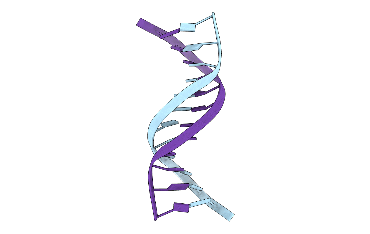
132D image
Deposition Date 1993-06-24
Release Date 1994-01-31
Last Version Date 2024-05-22
Entry Detail
PDB ID:
132D
Keywords:
Title:
SOLUTION STRUCTURE OF THE TN AN DNA DUPLEX GCCGTTAACGGC CONTAINING THE HPA I RESTRICTION SITE
Biological Source:
Source Organism:
(Taxon ID: )
Method Details:
Experimental Method:
Conformers Calculated:
1
Conformers Submitted:
1
Macromolecular Entities
Polymer Type:polydeoxyribonucleotide
Molecule:DNA (5'-D(P*GP*CP*CP*GP*TP*TP*AP*AP*CP*GP*GP*C)-3')
Chain IDs:A, B
Chain Length:12
Number of Molecules:2
Biological Source:
Ligand Molecules
Primary Citation
Solution structure of the TnAn DNA duplex GCCGTTAACGCG containing the HpaI restriction site.
Biochemistry 31 12103 12116 (1992)
PMID: 1457407 DOI: 10.1021/bi00163a020

Abstact

The solution structure of the self-complementary DNA duplex [d(GCCGTTAACGGC)]2, which contains the HpaI restriction site GTTAAC, has been elucidated by two-dimensional NMR, distance geometry (DG), and NOE back-calculation methods. Initial distance constraints were determined by polynomial fitting the two-spin initial NOE rates; backbone constraints from NOE and J-coupling observations (Kim et al., 1992) were included. RMSDs between initial-distance-refined structures derived from random-embedded DG, A-DNA, and B-DNA starting structures were all in the range 0.5-1.0 A, indicating good convergence properties of the algorithm, regardless of the starting structure. A semiautomatic back-calculation refinement procedure was developed and used to generate more refined structures for which the BKCALC-simulated NOE volumes matched the experimental data. The six final structures refined from various starting structures exhibit very good agreement with the experimental data (R values = 0.18) and converge well to within 0.8-A RMSD differences for the central 8 base pairs. The torsion and pseudorotation phase angles were found to be well determined by the data, and the local helical parameters for each base step converged quite well. The final structures show that the central T6-A7 step is somewhat underwound (twist angle ca. 29 degrees), with a large negative cup and a normal (wide) minor groove width, while the T5-T6 and A7-A8 steps have a partially narrowed minor groove.

Legend

Protein

Chemical

Disease

Primary Citation of related structures