11AS image
Deposition Date 1997-12-02
Release Date 1998-12-30
Last Version Date 2024-03-13
Entry Detail
PDB ID:
11AS
Keywords:
Title:
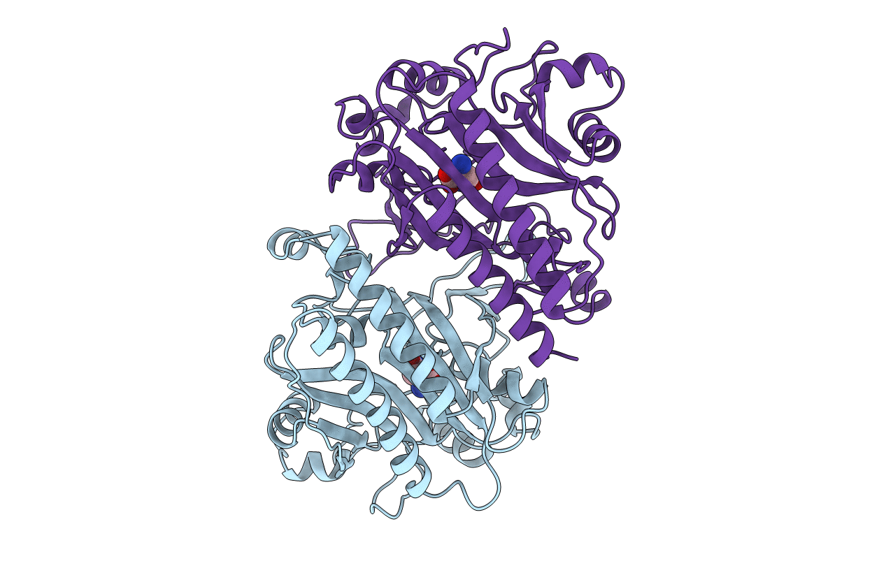
ASPARAGINE SYNTHETASE MUTANT C51A, C315A COMPLEXED WITH L-ASPARAGINE
Biological Source:
Source Organism:
Host Organism:
Method Details:
Experimental Method:
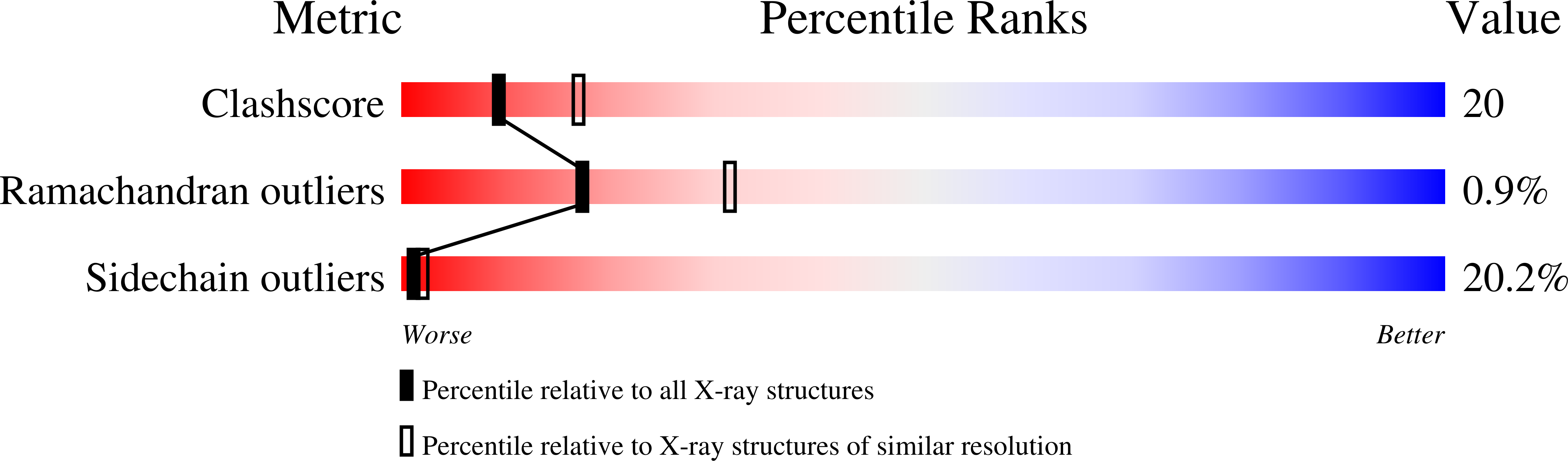
Resolution:
2.50 Å
R-Value Free:
0.25
R-Value Work:
0.15
R-Value Observed:
0.15
Space Group:
P 1 21 1
Macromolecular Entities
Structures with similar UniProt ID
Protein Blast
Polymer Type:polypeptide(L)
Molecule:ASPARAGINE SYNTHETASE
Gene (Uniprot):asnA
Mutagens:C51A, C315A
Chain IDs:A, B
Chain Length:330
Number of Molecules:2
Biological Source:Escherichia coli K12
Ligand Molecules
Primary Citation
Crystal structure of asparagine synthetase reveals a close evolutionary relationship to class II aminoacyl-tRNA synthetase.
Nat.Struct.Biol. 5 15 19 (1998)
PMID: 9437423 DOI: 10.1038/nsb0198-15

Abstact

The crystal structure of E. coli asparagine synthetase has been determined by X-ray diffraction analysis at 2.5 A resolution. The overall structure of the enzyme is remarkably similar to that of the catalytic domain of yeast aspartyl-tRNA synthetase despite low sequence similarity. These enzymes have a common reaction mechanism that implies the formation of an aminoacyl-adenylate intermediate. The active site architecture and most of the catalytic residues are also conserved in both enzymes. These proteins have probably evolved from a common ancestor even though their sequence similarities are small. The functional and structural similarities of both enzymes suggest that new enzymatic activities would generally follow the recruitment of a protein catalyzing a similar chemical reaction.

Legend

Protein

Chemical

Disease

Primary Citation of related structures
Feedback Form
Name
Email
Institute
Feedback