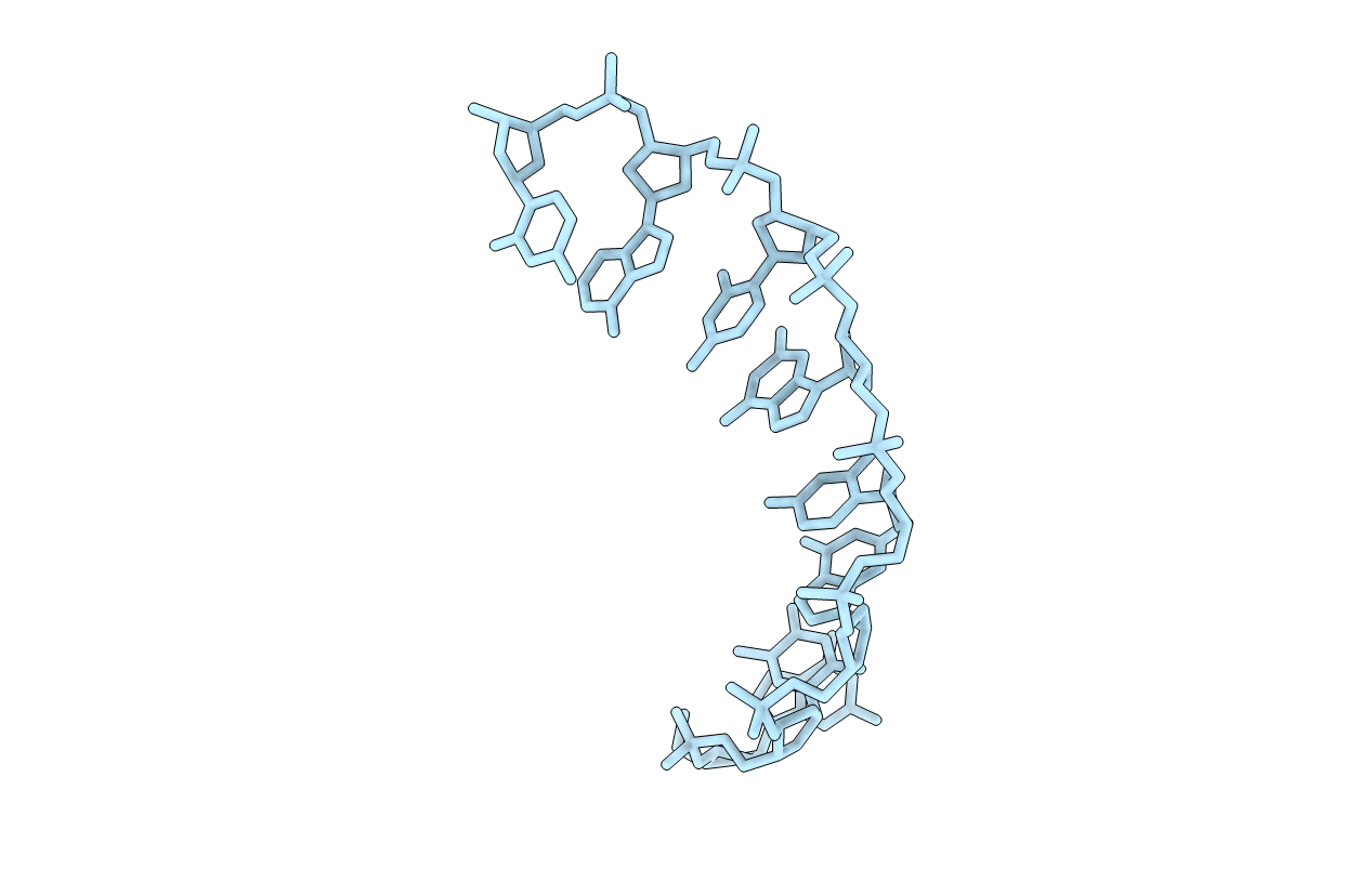
118D image
Deposition Date 1993-02-11
Release Date 1993-02-11
Last Version Date 2023-03-22
Entry Detail
PDB ID:
118D
Keywords:
Title:
CRYSTAL AND MOLECULAR STRUCTURE OF D(GTGCGCAC): INVESTIGATION OF THE EFFECTS OF BASE SEQUENCE ON THE CONFORMATION OF OCTAMER DUPLEXES
Biological Source:
Source Organism:
Method Details:
Experimental Method:
Resolution:
1.64 Å
R-Value Observed:
0.15
Space Group:
P 43 21 2
Macromolecular Entities
Polymer Type:polydeoxyribonucleotide
Molecule:DNA (5'-D(*GP*TP*GP*CP*GP*CP*AP*C)-3')
Chain IDs:A
Chain Length:8
Number of Molecules:1
Biological Source:
Primary Citation
Crystal and molecular structure of d(GTGCGCAC): investigation of the effects of base sequence on the conformation of octamer duplexes.
Biochemistry 31 12803 12812 (1992)
PMID: 1463751 DOI: 10.1021/bi00166a014

Abstact

The structure of the self-complementary deoxyoctanucleotide d(GTGCGCAC), which crystallized as an A-type helix in the space group P4(3)2(1)2, with one strand in the crystallographic asymmetric unit has been determined and refined to a final R-value of 0.154 using 1.64-A diffraction data collected on an area detector. In contrast to the closely related sequence d(GTGTACAC)tet, there was no evidence for an ordered spermine molecule in the major groove of this octamer. Ordered water is found associated with almost all the exposed hydrogen bonding groups of the octamer. A pentagonal ring of water molecules is hydrogen bonded to O6 and N7 of G3 and the N4 and O6 of the C4.G13 base pair. A detailed comparison of the local helical parameters of d(GTGCGCAC) and d(GTGTACAC)tet is presented. The base sequence change at the center of the octamers affects several of the local helical parameters, via both intra- and interduplex interactions within the crystal.

Legend

Protein

Chemical

Disease

Primary Citation of related structures