
Search Count: 25
 |
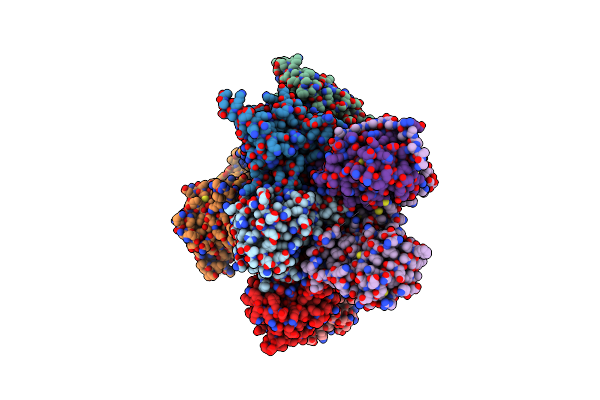
Structure Of The Non-Canonical Ctlh E3 Substrate Receptor Wdr26 Bound To Ypel5
Organism: Homo sapiens
Method: ELECTRON MICROSCOPY Release Date: 2024-05-15 Classification: LIGASE Ligands: ZN |
 |
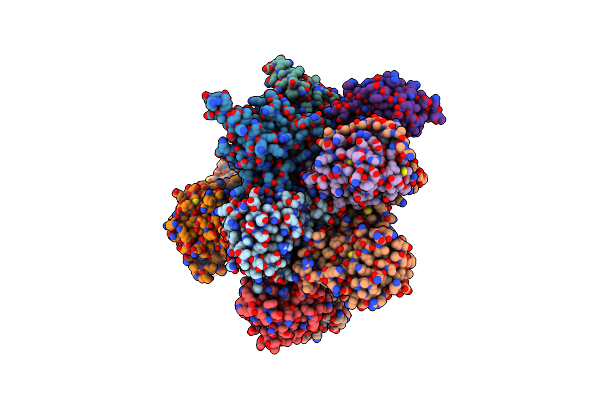
Structure Of The Non-Canonical Ctlh E3 Substrate Receptor Wdr26 Bound To Nmnat1 Substrate
Organism: Homo sapiens
Method: ELECTRON MICROSCOPY Release Date: 2024-05-15 Classification: LIGASE Ligands: NMN, ZN |
 |
Organism: Homo sapiens
Method: ELECTRON MICROSCOPY Release Date: 2024-02-21 Classification: HYDROLASE |
 |
Organism: Homo sapiens
Method: ELECTRON MICROSCOPY Release Date: 2024-02-21 Classification: HYDROLASE |
 |
Organism: Homo sapiens
Method: ELECTRON MICROSCOPY Release Date: 2024-02-21 Classification: HYDROLASE |
 |
Organism: Homo sapiens
Method: ELECTRON MICROSCOPY Release Date: 2024-02-21 Classification: HYDROLASE |
 |
Organism: Homo sapiens
Method: ELECTRON MICROSCOPY Release Date: 2024-02-21 Classification: HYDROLASE |
 |
Organism: Homo sapiens
Method: ELECTRON MICROSCOPY Release Date: 2024-02-21 Classification: HYDROLASE |
 |
Organism: Homo sapiens
Method: ELECTRON MICROSCOPY Release Date: 2024-02-21 Classification: HYDROLASE |
 |
Organism: Saccharomyces cerevisiae
Method: ELECTRON MICROSCOPY Release Date: 2024-01-17 Classification: LIGASE Ligands: PCW, PX6 |
 |
Organism: Saccharomyces cerevisiae
Method: ELECTRON MICROSCOPY Release Date: 2024-01-17 Classification: LIGASE Ligands: PCW, PX6 |
 |
Catalytic Module Of Human Ctlh E3 Ligase Bound To Multiphosphorylated Ube2H~Ubiquitin
Organism: Homo sapiens
Method: ELECTRON MICROSCOPY Release Date: 2024-01-03 Classification: LIGASE Ligands: ZN |
 |
Catalytic Module Of Yeast Gid E3 Ligase Bound To Multiphosphorylated Ubc8~Ubiquitin
Organism: Saccharomyces cerevisiae, Homo sapiens
Method: ELECTRON MICROSCOPY Release Date: 2024-01-03 Classification: LIGASE Ligands: ZN |
 |
Organism: Homo sapiens
Method: ELECTRON MICROSCOPY Release Date: 2023-12-13 Classification: ANTIVIRAL PROTEIN Ligands: AMP |
 |
Structure Of Crl7Fbxw8 Reveals Coupling With Cul1-Rbx1/Roc1 For Multi-Cullin-Ring E3-Catalyzed Ubiquitin Ligation
Organism: Homo sapiens
Method: ELECTRON MICROSCOPY Release Date: 2022-08-24 Classification: LIGASE Ligands: ZN |
 |
Organism: Saccharomyces cerevisiae yjm1133, Saccharomyces cerevisiae
Method: ELECTRON MICROSCOPY Release Date: 2022-06-08 Classification: LIGASE |
 |
Organism: Homo sapiens
Method: X-RAY DIFFRACTION Resolution:2.45 Å Release Date: 2021-09-15 Classification: LIGASE Ligands: ZN |
 |
Organism: Homo sapiens
Method: ELECTRON MICROSCOPY Release Date: 2021-09-15 Classification: LIGASE Ligands: ZN |
 |
Substrate Receptor Scaffolding Module Of Yeast Chelator-Gid Sr4 E3 Ubiquitin Ligase Bound To Fbp1 Substrate
Organism: Saccharomyces cerevisiae (strain atcc 204508 / s288c), Saccharomyces cerevisiae
Method: ELECTRON MICROSCOPY Release Date: 2021-05-05 Classification: LIGASE |
 |
Organism: Saccharomyces cerevisiae (strain atcc 204508 / s288c)
Method: ELECTRON MICROSCOPY Release Date: 2021-05-05 Classification: LIGASE Ligands: ZN |