
Search Count: 37
 |
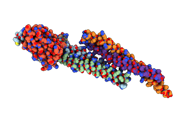
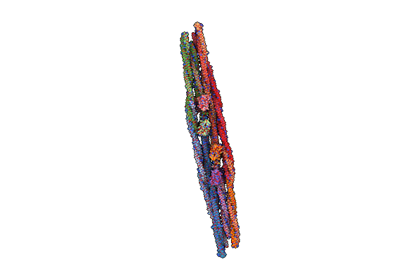
Cryo-Em Structure Of Crescentin Filaments (Stutter Mutant, C1 Symmetry And Small Box)
Organism: Caulobacter vibrioides, Camelidae
Method: ELECTRON MICROSCOPY Release Date: 2023-08-16 Classification: STRUCTURAL PROTEIN |
 |
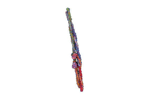
Cryo-Em Structure Of Crescentin Filaments (Stutter Mutant, C2, Symmetry And Small Box)
Organism: Caulobacter vibrioides, Camelidae
Method: ELECTRON MICROSCOPY Release Date: 2023-08-16 Classification: STRUCTURAL PROTEIN |
 |
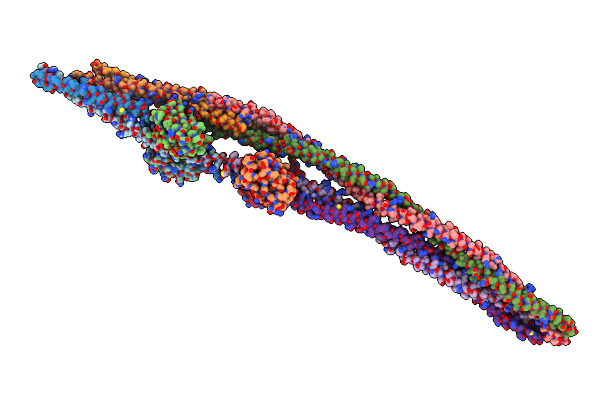
Cryo-Em Structure Of Crescentin Filaments (Wildtype, C1 Symmetry And Small Box)
Organism: Caulobacter vibrioides, Camelidae
Method: ELECTRON MICROSCOPY Release Date: 2023-08-16 Classification: STRUCTURAL PROTEIN |
 |
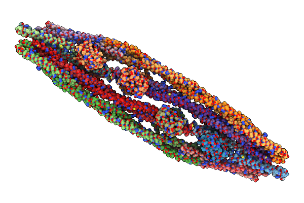
Cryo-Em Structure Of Crescentin Filaments (Wildtype, C2 Symmetry And Small Box)
Organism: Caulobacter vibrioides, Camelidae
Method: ELECTRON MICROSCOPY Release Date: 2023-08-16 Classification: STRUCTURAL PROTEIN |
 |
Cryo-Em Structure Of Crescentin Filaments (Stutter Mutant, C1 Symmetry And Large Box)
Organism: Caulobacter vibrioides, Camelidae
Method: ELECTRON MICROSCOPY Resolution:4.10 Å Release Date: 2023-08-16 Classification: STRUCTURAL PROTEIN |
 |
Cryo-Em Structure Of Crescentin Filaments (Wildtype, C1 Symmetry And Large Box)
Organism: Caulobacter vibrioides, Camelidae
Method: ELECTRON MICROSCOPY Release Date: 2023-08-16 Classification: STRUCTURAL PROTEIN |
 |
Cryo-Em Structure Of Crescentin Filaments (Wildtype, C2 Symmetry And Large Box)
Organism: Caulobacter vibrioides, Camelidae
Method: ELECTRON MICROSCOPY Release Date: 2023-08-16 Classification: STRUCTURAL PROTEIN |
 |
Cryo-Em Structure Of Crescentin Filaments (Stutter Mutant, C2 Symmetry And Large Box)
Organism: Caulobacter vibrioides, Camelidae
Method: ELECTRON MICROSCOPY Release Date: 2023-08-16 Classification: STRUCTURAL PROTEIN |
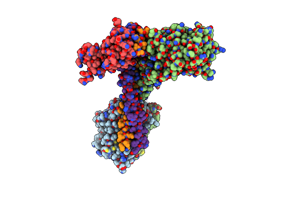 |
Organism: Pseudomonas aeruginosa pao1
Method: ELECTRON MICROSCOPY Release Date: 2023-04-19 Classification: MEMBRANE PROTEIN |
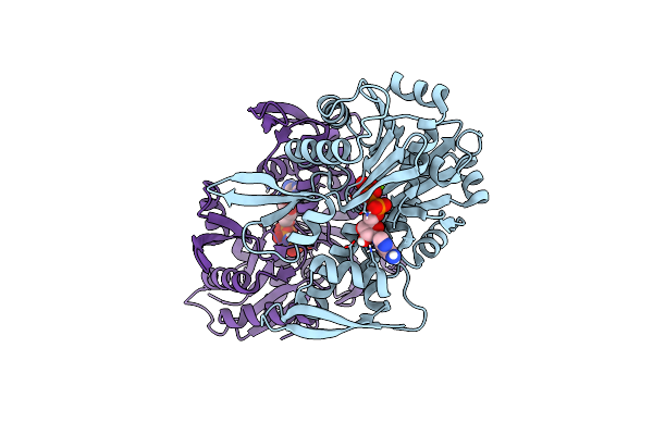 |
Organism: Caulobacter vibrioides
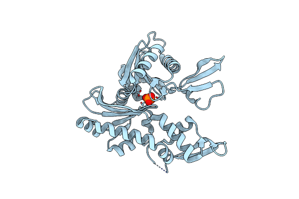
Method: X-RAY DIFFRACTION Resolution:2.20 Å Release Date: 2014-08-13 Classification: STRUCTURAL PROTEIN Ligands: MG, ANP |
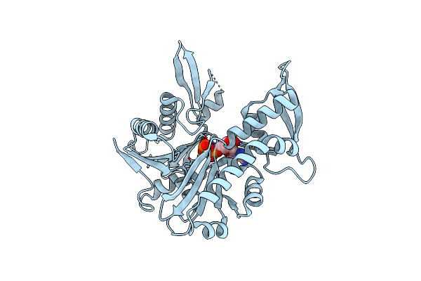 |
Organism: Caulobacter vibrioides
Method: X-RAY DIFFRACTION Resolution:1.60 Å Release Date: 2014-06-25 Classification: STRUCTURAL PROTEIN Ligands: MG, ADP |
 |
Organism: Caulobacter vibrioides
Method: X-RAY DIFFRACTION Resolution:2.00 Å Release Date: 2014-06-11 Classification: STRUCTURAL PROTEIN Ligands: PO4 |
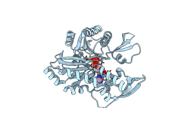 |
Organism: Caulobacter vibrioides
Method: X-RAY DIFFRACTION Resolution:1.64 Å Release Date: 2014-06-11 Classification: STRUCTURAL PROTEIN Ligands: MG, ADP |
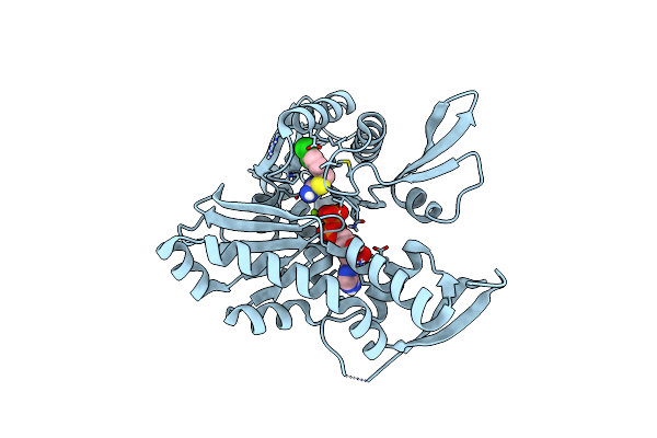 |
Organism: Caulobacter vibrioides
Method: X-RAY DIFFRACTION Resolution:1.50 Å Release Date: 2014-06-11 Classification: STRUCTURAL PROTEIN Ligands: QH3, ADP, MG |
 |
Organism: Caulobacter vibrioides
Method: X-RAY DIFFRACTION Resolution:1.64 Å Release Date: 2014-06-11 Classification: STRUCTURAL PROTEIN Ligands: F90, ADP, MG |
 |
Organism: Caulobacter vibrioides
Method: X-RAY DIFFRACTION Resolution:1.80 Å Release Date: 2014-06-11 Classification: STRUCTURAL PROTEIN |
 |
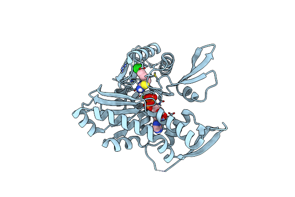
Organism: Caulobacter vibrioides
Method: X-RAY DIFFRACTION Resolution:2.00 Å Release Date: 2014-06-11 Classification: STRUCTURAL PROTEIN Ligands: MG, ANP |
 |
Organism: Caulobacter vibrioides
Method: X-RAY DIFFRACTION Resolution:2.60 Å Release Date: 2014-06-11 Classification: STRUCTURAL PROTEIN Ligands: MG, ANP, F90 |
 |
Organism: Escherichia coli
Method: ELECTRON MICROSCOPY Resolution:7.20 Å Release Date: 2012-11-21 Classification: TRANSPORT PROTEIN Ligands: ANP, MG |
 |
Organism: Escherichia coli
Method: X-RAY DIFFRACTION Resolution:2.00 Å Release Date: 2012-11-07 Classification: TRANSPORT PROTEIN Ligands: ANP, MG |